1BZH

Cyclic peptide inhibitor of human PTP1B


Experimental Data Snapshot

  • Method: X-RAY DIFFRACTION
  • Resolution: 2.10 Å
  • R-Value Free: 0.262 
  • R-Value Work: 0.196 

wwPDB Validation   3D Report Full Report


This is version 2.2 of the entry. See complete history


Literature

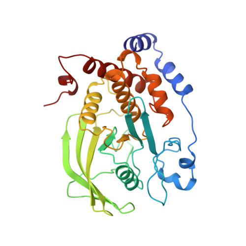
Structural basis for inhibition of the protein tyrosine phosphatase 1B by phosphotyrosine peptide mimetics.

Groves, M.R.Yao, Z.J.Roller, P.P.Burke Jr., T.R.Barford, D.

(1998) Biochemistry 37: 17773-17783

  • DOI: https://doi.org/10.1021/bi9816958
  • Primary Citation of Related Structures:  
    1BZC, 1BZH, 1BZJ

  • PubMed Abstract: 

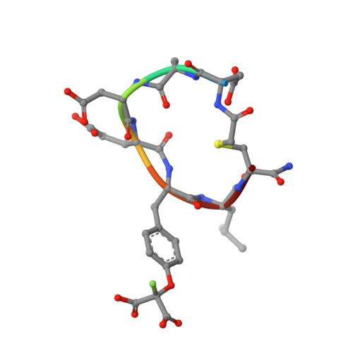
    Protein tyrosine phosphatases regulate diverse cellular processes and represent important targets for therapeutic intervention in a number of diseases. The crystal structures of protein tyrosine phosphatase 1B (PTP1B) in complex with small molecule inhibitors based upon two classes of phosphotyrosine mimetics, the (difluoronaphthylmethyl)phosphonic acids and the fluoromalonyl tyrosines, have been determined to resolutions greater than 2.3 A. The fluoromalonyl tyrosine residue was incorporated within a cyclic hexapeptide modeled on an autophosphorylation site of the epidermal growth factor receptor. The structure of this inhibitor bound to PTP1B represents the first crystal structure of a non-phosphonate-containing inhibitor and reveals the mechanism of phosphotyrosine mimicry by the fluoromalonyl tyrosine residue and the nature of its interactions within the catalytic site of PTP1B. In contrast to complexes of PTP1B with phosphotyrosine-containing peptides, binding of the fluoromalonyl tyrosine residue to the catalytic site of PTP1B is not accompanied by closure of the catalytic site WPD loop. Structures of PTP1B in complex with the (difluoronaphthylmethyl)phosphonic acid derivatives reveal that substitutions of the naphthalene ring modulate the mode of inhibitor binding to the catalytic site and provide the potential for enhanced inhibitor affinity and the generation of PTP-specific inhibitors. These results provide a framework for the rational design of higher affinity and more specific phosphotyrosine mimetic inhibitors of not only protein tyrosine phosphatases but also SH2 and PTB domains.


  • Organizational Affiliation

    Department of Biochemistry, University of Oxford, United Kingdom.


Macromolecules
Find similar proteins by:  (by identity cutoff)  |  3D Structure
Entity ID: 1
MoleculeChains Sequence LengthOrganismDetailsImage
PROTEIN (PROTEIN-TYROSINE-PHOSPHATASE 1B)298Homo sapiensMutation(s): 0 
EC: 3.1.3.48
UniProt & NIH Common Fund Data Resources
Find proteins for P18031 (Homo sapiens)
Explore P18031 
Go to UniProtKB:  P18031
PHAROS:  P18031
GTEx:  ENSG00000196396 
Entity Groups  
Sequence Clusters30% Identity50% Identity70% Identity90% Identity95% Identity100% Identity
UniProt GroupP18031
Sequence Annotations
Expand
  • Reference Sequence

Find similar proteins by:  Sequence   |   3D Structure  

Entity ID: 2
MoleculeChains Sequence LengthOrganismDetailsImage
PROTEIN (PROTEIN-TYROSINE-PHOSPHATASE 1B INHIBITOR)B [auth I]7Homo sapiensMutation(s): 0 
Sequence Annotations
Expand
  • Reference Sequence
Small Molecules
Modified Residues  2 Unique
IDChains TypeFormula2D DiagramParent
AEA
Query on AEA
B [auth I]L-PEPTIDE LINKINGC5 H10 N2 O3 SCYS
FLT
Query on FLT
B [auth I]L-PEPTIDE LINKINGC12 H12 F N O7TYR
Experimental Data & Validation

Experimental Data

  • Method: X-RAY DIFFRACTION
  • Resolution: 2.10 Å
  • R-Value Free: 0.262 
  • R-Value Work: 0.196 
  • Space Group: P 1 21 1
Unit Cell:
Length ( Å )Angle ( ˚ )
a = 39.69α = 90
b = 86.82β = 96.91
c = 52γ = 90
Software Package:
Software NamePurpose
AMoREphasing
REFMACrefinement
DENZOdata reduction
CCP4data scaling

Structure Validation

View Full Validation Report


Entry History 

Deposition Data

Revision History  (Full details and data files)

  • Version 1.0: 1999-02-16
    Type: Initial release
  • Version 1.1: 2008-04-27
    Changes: Version format compliance
  • Version 1.2: 2011-07-13
    Changes: Atomic model, Database references, Derived calculations, Non-polymer description, Structure summary, Version format compliance
  • Version 2.0: 2018-12-26
    Changes: Data collection, Polymer sequence
  • Version 2.1: 2023-08-09
    Changes: Data collection, Database references, Derived calculations, Refinement description
  • Version 2.2: 2023-11-15
    Changes: Data collection