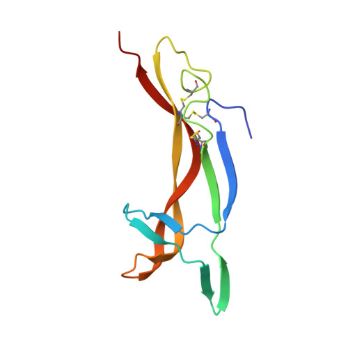
Nerve growth factor in different crystal forms displays structural flexibility and reveals zinc binding sites.
Holland, D.R., Cousens, L.S., Meng, W., Matthews, B.W.(1994) J Mol Biol 239: 385-400
- PubMed: 8201620
- DOI: https://doi.org/10.1006/jmbi.1994.1380
- Primary Citation of Related Structures:
1BTG - PubMed Abstract:
Murine beta-nerve growth factor (beta NGF) is a 118 amino acid residue polypeptide which, as a functional dimer, plays an important role in the survival and development of certain neuronal populations. The structure of the bis-desocta1-8 form of murine beta NGF has been determined in two different crystal modifications using X-ray methods. The two crystal forms, with space groups P2(1)2(1)2(1) and C2, were grown from 18 to 20% polyethylene glycol 8000 and 100 mM Pipes (pH 6.1) with zinc acetate concentrations of 1 mM and 100 mM, respectively. The C2 structure was solved by multiple isomorphous replacement using four heavy-atom derivatives and was refined to a crystallographic residual of 17.9% and 2.5 A resolution. The crystals contain three beta NGF monomers per asymmetric unit. Two monomers form a dimer related by a non-crystallographic 2-fold axis of symmetry. The third monomer also forms a dimer that is very similar, but with a crystallography related monomer as a partner. The electron density clearly defines residues 12 through 115 for all three monomers but the extreme N and C-terminal residues (9 to 11, 116 to 118) are ill defined in some cases. The P2(1)2(1)2(1) structure was solved by molecular replacement using the C2 structure as a search model and was refined to a crystallographic residual of 19.7% at 2.8 A resolution. This crystal form contains two monomers per asymmetric unit, again arranged as a non-crystallographic 2-fold-related dimer. The N and c termini are also variably defined. The core of each of the five monomers, which forms a cysteine knot motif, is very similar in all structures. Also, the dimer structures are very similar to one another, whether the monomers are related by crystallographic or non-crystallographic symmetry. However, three of the four loop regions that extend from the core of each monomer display substantial variability in conformation, even between monomers of the same dimer. This structural variability in the putative receptor binding regions suggests that structural malleability might be important in allowing the ligands to bind to different receptors with different affinities.(ABSTRACT TRUNCATED AT 400 WORDS)
Organizational Affiliation:
Institute of Molecular Biology, Howard Hughes Medical Institute, Eugene, OR.