1BRL

THREE-DIMENSIONAL STRUCTURE OF BACTERIAL LUCIFERASE FROM VIBRIO HARVEYI AT 2.4 ANGSTROMS RESOLUTION


Experimental Data Snapshot

  • Method: X-RAY DIFFRACTION
  • Resolution: 2.40 Å
  • R-Value Observed: 0.208 

wwPDB Validation   3D Report Full Report


This is version 1.3 of the entry. See complete history


Literature

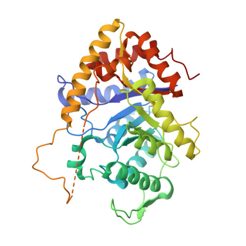
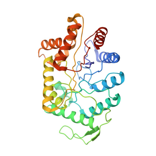
Three-dimensional structure of bacterial luciferase from Vibrio harveyi at 2.4 A resolution.

Fisher, A.J.Raushel, F.M.Baldwin, T.O.Rayment, I.

(1995) Biochemistry 34: 6581-6586

  • DOI: https://doi.org/10.1021/bi00020a002
  • Primary Citation of Related Structures:  
    1BRL

  • PubMed Abstract: 

    Luciferases are a class of enzymes that generate light in the visible spectrum. Luciferase from luminous marine bacteria is an alpha-beta heterodimer monooxygenase that catalyzes the oxidation of FMNH2 and a long-chain aliphatic aldehyde. The X-ray crystal structure of bacterial luciferase from Vibrio harveyi has been determined to 2.4 A resolution. The structure was solved by a combination of multiple isomorphous replacement and molecular averaging between the two heterodimers in the asymmetric unit. Each subunit folds into a (beta/alpha)8 barrel motif, and dimerization is mediated through a parallel four-helix bundle centered on a pseudo 2-fold axis that relates the structurally similar subunits. The vicinity of the active site has been identified on the alpha subunit by correlations with similar protein motifs and previous biochemical studies. The structure presented here represents the first molecular model of a bioluminescent enzyme.


  • Organizational Affiliation

    Department of Biochemistry, University of Wisconsin, Madison 53705, USA.


Macromolecules
Find similar proteins by:  (by identity cutoff)  |  3D Structure
Entity ID: 1
MoleculeChains Sequence LengthOrganismDetailsImage
BACTERIAL LUCIFERASE
A, C
355Vibrio harveyiMutation(s): 0 
EC: 1.14.14.3
UniProt
Find proteins for P07740 (Vibrio harveyi)
Explore P07740 
Go to UniProtKB:  P07740
Entity Groups  
Sequence Clusters30% Identity50% Identity70% Identity90% Identity95% Identity100% Identity
UniProt GroupP07740
Sequence Annotations
Expand
  • Reference Sequence
Find similar proteins by:  (by identity cutoff)  |  3D Structure
Entity ID: 2
MoleculeChains Sequence LengthOrganismDetailsImage
BACTERIAL LUCIFERASE
B, D
324Vibrio harveyiMutation(s): 0 
EC: 1.14.14.3
UniProt
Find proteins for P07739 (Vibrio harveyi)
Explore P07739 
Go to UniProtKB:  P07739
Entity Groups  
Sequence Clusters30% Identity50% Identity70% Identity90% Identity95% Identity100% Identity
UniProt GroupP07739
Sequence Annotations
Expand
  • Reference Sequence
Small Molecules
Ligands 1 Unique
IDChains Name / Formula / InChI Key2D Diagram3D Interactions
PO4
Query on PO4

Download Ideal Coordinates CCD File 
E [auth A]PHOSPHATE ION
O4 P
NBIIXXVUZAFLBC-UHFFFAOYSA-K
Experimental Data & Validation

Experimental Data

  • Method: X-RAY DIFFRACTION
  • Resolution: 2.40 Å
  • R-Value Observed: 0.208 
  • Space Group: P 21 21 21
Unit Cell:
Length ( Å )Angle ( ˚ )
a = 59.9α = 90
b = 112.7β = 90
c = 301.8γ = 90
Software Package:
Software NamePurpose
TNTrefinement
PURDUEdata reduction

Structure Validation

View Full Validation Report


Entry History 

Deposition Data

Revision History  (Full details and data files)

  • Version 1.0: 1996-04-01
    Type: Initial release
  • Version 1.1: 2008-03-24
    Changes: Version format compliance
  • Version 1.2: 2011-07-13
    Changes: Version format compliance
  • Version 1.3: 2024-02-07
    Changes: Data collection, Database references, Derived calculations, Other