1BRB

CRYSTAL STRUCTURES OF RAT ANIONIC TRYPSIN COMPLEXED WITH THE PROTEIN INHIBITORS APPI AND BPTI


Experimental Data Snapshot

  • Method: X-RAY DIFFRACTION
  • Resolution: 2.10 Å
  • R-Value Work: 0.198 
  • R-Value Observed: 0.198 

wwPDB Validation   3D Report Full Report


This is version 1.3 of the entry. See complete history


Literature

Crystal structures of rat anionic trypsin complexed with the protein inhibitors APPI and BPTI.

Perona, J.J.Tsu, C.A.Craik, C.S.Fletterick, R.J.

(1993) J Mol Biol 230: 919-933

  • DOI: https://doi.org/10.1006/jmbi.1993.1210
  • Primary Citation of Related Structures:  
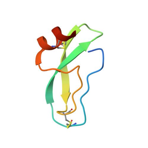
    1BRB

  • PubMed Abstract: 

    The crystal structure of rat anionic trypsin D189G/G226D has been determined in complexes with each of the protein inhibitors APPI (amyloid beta-protein precursor inhibitor domain) and BPTI (bovine pancreatic trypsin inhibitor) at resolutions of 2.5 A and 2.1 A, respectively. Comparisons with the structure of the bovine trypsin-BPTI complex show that the enzyme-inhibitor interactions in rat trypsin are dominated to a much greater degree by attractive and repulsive electrostatic forces. Decreased structural complementarity in the flanking regions of the interface formed with BPTI is reflected in significantly weaker inhibition relative to bovine trypsin. The primary active site loop of BPTI adopts slightly different conformations when bound to rat and cow trypsins, reflecting a broader entrance to the binding pocket in the former. Tight complementarity of each loop conformer to the respective active sites then gives rise to significantly different overall orientations of the inhibitor when bound to the two enzymes. The crystal structures of trypsin bound to these protein inhibitors are excellent models of the Michaelis complexes, which permit visualization of substrate interactions both N and C-terminal to the cleaved bond, while maintaining identical reaction chemistry. They will be uniquely useful to the structure-function analysis of variant rat trypsin enzymes.


  • Organizational Affiliation

    Department of Pharmaceutical Chemistry, University of California, San Francisco 94143-0446.


Macromolecules
Find similar proteins by:  (by identity cutoff)  |  3D Structure
Entity ID: 1
MoleculeChains Sequence LengthOrganismDetailsImage
TRYPSINA [auth E]223Rattus norvegicusMutation(s): 0 
EC: 3.4.21.4
UniProt
Find proteins for P00763 (Rattus norvegicus)
Explore P00763 
Go to UniProtKB:  P00763
Entity Groups  
Sequence Clusters30% Identity50% Identity70% Identity90% Identity95% Identity100% Identity
UniProt GroupP00763
Sequence Annotations
Expand
  • Reference Sequence
Find similar proteins by:  (by identity cutoff)  |  3D Structure
Entity ID: 2
MoleculeChains Sequence LengthOrganismDetailsImage
PANCREATIC TRYPSIN INHIBITORB [auth I]58Bos taurusMutation(s): 0 
UniProt
Find proteins for P00974 (Bos taurus)
Explore P00974 
Go to UniProtKB:  P00974
Entity Groups  
Sequence Clusters30% Identity50% Identity70% Identity90% Identity95% Identity100% Identity
UniProt GroupP00974
Sequence Annotations
Expand
  • Reference Sequence
Experimental Data & Validation

Experimental Data

  • Method: X-RAY DIFFRACTION
  • Resolution: 2.10 Å
  • R-Value Work: 0.198 
  • R-Value Observed: 0.198 
  • Space Group: P 32 2 1
Unit Cell:
Length ( Å )Angle ( ˚ )
a = 92.65α = 90
b = 92.65β = 90
c = 62.32γ = 120
Software Package:
Software NamePurpose
X-PLORmodel building
X-PLORrefinement
X-PLORphasing

Structure Validation

View Full Validation Report


Entry History 

Deposition Data

Revision History  (Full details and data files)

  • Version 1.0: 1994-07-31
    Type: Initial release
  • Version 1.1: 2008-03-03
    Changes: Version format compliance
  • Version 1.2: 2011-07-13
    Changes: Version format compliance
  • Version 1.3: 2017-11-29
    Changes: Derived calculations, Other