1BOW

MULTIDRUG-BINDING DOMAIN OF TRANSCRIPTION ACTIVATOR BMRR (APO FORM)


Experimental Data Snapshot

  • Method: X-RAY DIFFRACTION
  • Resolution: 2.70 Å
  • R-Value Free: 0.270 
  • R-Value Observed: 0.202 

wwPDB Validation   3D Report Full Report


This is version 1.3 of the entry. See complete history


Literature

Structural basis of multidrug recognition by BmrR, a transcription activator of a multidrug transporter.

Zheleznova, E.E.Markham, P.N.Neyfakh, A.A.Brennan, R.G.

(1999) Cell 96: 353-362

  • DOI: https://doi.org/10.1016/s0092-8674(00)80548-6
  • Primary Citation of Related Structures:  
    1BOW, 2BOW

  • PubMed Abstract: 

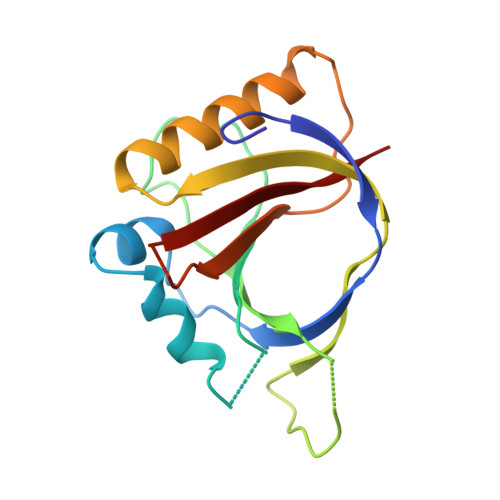
    Multidrug-efflux transporters demonstrate an unusual ability to recognize multiple structurally dissimilar toxins. A comparable ability to bind diverse hydrophobic cationic drugs is characteristic of the Bacillus subtilis transcription regulator BmrR, which upon drug binding activates expression of the multidrug transporter Bmr. Crystal structures of the multidrug-binding domain of BmrR (2.7 A resolution) and of its complex with the drug tetraphenylphosphonium (2.8 A resolution) revealed a drug-induced unfolding and relocation of an alpha helix, which exposes an internal drug-binding pocket. Tetraphenylphosphonium binding is mediated by stacking and van der Waals contacts with multiple hydrophobic residues of the pocket and by an electrostatic interaction between the positively charged drug and a buried glutamate residue, which is the key to cation selectivity. Similar binding principles may be used by other multidrug-binding proteins.


  • Organizational Affiliation

    Department of Biochemistry and Molecular Biology, Oregon Health Sciences University, Portland 97201-3098, USA.


Macromolecules
Find similar proteins by:  (by identity cutoff)  |  3D Structure
Entity ID: 1
MoleculeChains Sequence LengthOrganismDetailsImage
MULTIDRUG-EFFLUX TRANSPORTER 1 REGULATOR BMRR159Bacillus subtilisMutation(s): 0 
UniProt
Find proteins for P39075 (Bacillus subtilis (strain 168))
Explore P39075 
Go to UniProtKB:  P39075
Entity Groups  
Sequence Clusters30% Identity50% Identity70% Identity90% Identity95% Identity100% Identity
UniProt GroupP39075
Sequence Annotations
Expand
  • Reference Sequence
Small Molecules
Ligands 1 Unique
IDChains Name / Formula / InChI Key2D Diagram3D Interactions
MN
Query on MN

Download Ideal Coordinates CCD File 
B [auth A]MANGANESE (II) ION
Mn
WAEMQWOKJMHJLA-UHFFFAOYSA-N
Experimental Data & Validation

Experimental Data

  • Method: X-RAY DIFFRACTION
  • Resolution: 2.70 Å
  • R-Value Free: 0.270 
  • R-Value Observed: 0.202 
  • Space Group: P 41 21 2
Unit Cell:
Length ( Å )Angle ( ˚ )
a = 76.3α = 90
b = 76.3β = 90
c = 96γ = 90
Software Package:
Software NamePurpose
PHASESphasing
TNTrefinement
ADSCdata collection
LOSCALEdata scaling

Structure Validation

View Full Validation Report


Entry History 

Deposition Data

Revision History  (Full details and data files)

  • Version 1.0: 1999-08-06
    Type: Initial release
  • Version 1.1: 2008-03-03
    Changes: Version format compliance
  • Version 1.2: 2011-07-13
    Changes: Version format compliance
  • Version 1.3: 2024-02-07
    Changes: Data collection, Database references, Derived calculations