1BOB

HISTONE ACETYLTRANSFERASE HAT1 FROM SACCHAROMYCES CEREVISIAE IN COMPLEX WITH ACETYL COENZYME A


Experimental Data Snapshot

  • Method: X-RAY DIFFRACTION
  • Resolution: 2.30 Å
  • R-Value Free: 0.269 
  • R-Value Work: 0.204 
  • R-Value Observed: 0.204 

wwPDB Validation   3D Report Full Report


Ligand Structure Quality Assessment 


This is version 1.3 of the entry. See complete history


Literature

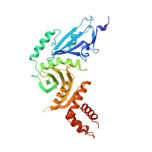
Structure of the histone acetyltransferase Hat1: a paradigm for the GCN5-related N-acetyltransferase superfamily.

Dutnall, R.N.Tafrov, S.T.Sternglanz, R.Ramakrishnan, V.

(1998) Cell 94: 427-438

  • DOI: https://doi.org/10.1016/s0092-8674(00)81584-6
  • Primary Citation of Related Structures:  
    1BOB

  • PubMed Abstract: 

    We have solved the crystal structure of the yeast histone acetyltransferase Hat1-acetyl coenzyme A (AcCoA) complex at 2.3 A resolution. Hat1 has an elongated, curved structure, and the AcCoA molecule is bound in a cleft on the concave surface of the protein, marking the active site of the enzyme. A channel of variable width and depth that runs across the protein is probably the binding site for the histone substrate. A model for histone H4 binding by Hat1 is discussed in terms of possible sources of specific lysine recognition by the enzyme. The structure of Hat1 provides a model for the structures of the catalytic domains of a protein superfamily that includes other histone acetyltransferases such as Gcn5 and CBP.


  • Organizational Affiliation

    Department of Biochemistry, University of Utah School of Medicine, Salt Lake City 84132, USA. rnd@snowbird.med.utah.edu


Macromolecules
Find similar proteins by:  (by identity cutoff)  |  3D Structure
Entity ID: 1
MoleculeChains Sequence LengthOrganismDetailsImage
HISTONE ACETYLTRANSFERASE320Saccharomyces cerevisiaeMutation(s): 0 
Gene Names: HAT1
EC: 2.3.1.48
UniProt
Find proteins for Q12341 (Saccharomyces cerevisiae (strain ATCC 204508 / S288c))
Explore Q12341 
Go to UniProtKB:  Q12341
Entity Groups  
Sequence Clusters30% Identity50% Identity70% Identity90% Identity95% Identity100% Identity
UniProt GroupQ12341
Sequence Annotations
Expand
  • Reference Sequence
Small Molecules
Ligands 2 Unique
IDChains Name / Formula / InChI Key2D Diagram3D Interactions
ACO
Query on ACO

Download Ideal Coordinates CCD File 
C [auth A]ACETYL COENZYME *A
C23 H38 N7 O17 P3 S
ZSLZBFCDCINBPY-ZSJPKINUSA-N
CA
Query on CA

Download Ideal Coordinates CCD File 
B [auth A]CALCIUM ION
Ca
BHPQYMZQTOCNFJ-UHFFFAOYSA-N
Experimental Data & Validation

Experimental Data

  • Method: X-RAY DIFFRACTION
  • Resolution: 2.30 Å
  • R-Value Free: 0.269 
  • R-Value Work: 0.204 
  • R-Value Observed: 0.204 
  • Space Group: C 1 2 1
Unit Cell:
Length ( Å )Angle ( ˚ )
a = 111.3α = 90
b = 47.9β = 90.3
c = 75.7γ = 90
Software Package:
Software NamePurpose
SOLVEphasing
X-PLORmodel building
X-PLORrefinement
DENZOdata reduction
SCALEPACKdata scaling
X-PLORphasing

Structure Validation

View Full Validation Report


Ligand Structure Quality Assessment 


Entry History 

Deposition Data

Revision History  (Full details and data files)

  • Version 1.0: 1999-04-20
    Type: Initial release
  • Version 1.1: 2008-03-24
    Changes: Version format compliance
  • Version 1.2: 2011-07-13
    Changes: Version format compliance
  • Version 1.3: 2024-02-07
    Changes: Data collection, Database references, Derived calculations