Nucleoside diphosphate kinase from bovine retina: purification, subcellular localization, molecular cloning, and three-dimensional structure.
Abdulaev, N.G., Karaschuk, G.N., Ladner, J.E., Kakuev, D.L., Yakhyaev, A.V., Tordova, M., Gaidarov, I.O., Popov, V.I., Fujiwara, J.H., Chinchilla, D., Eisenstein, E., Gilliland, G.L., Ridge, K.D.(1998) Biochemistry 37: 13958-13967
- PubMed: 9760230
- DOI: https://doi.org/10.1021/bi980853s
- Primary Citation of Related Structures:
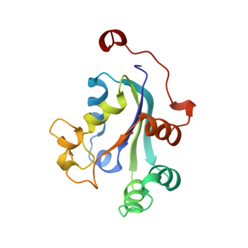
1BE4 - PubMed Abstract:
The biochemical and structural properties of bovine retinal nucleoside diphosphate kinase were investigated. The enzyme showed two polypeptides of approximately 17.5 and 18.5 kDa on SDS-PAGE, while isoelectric focusing revealed seven to eight proteins with a pI range of 7.4-8.2. Sedimentation equilibrium yielded a molecular mass of 96 +/- 2 kDa for the enzyme. Carbohydrate analysis revealed that both polypeptides contained Gal, Man, GlcNAc, Fuc, and GalNac saccharides. Like other nucleoside diphosphate kinases, the retinal enzyme showed substantial differences in the Km values for various di- and triphosphate nucleotides. Immunogold labeling of bovine retina revealed that the enzyme is localized on both the membranes and in the cytoplasm. Screening of a retinal cDNA library yielded full-length clones encoding two distinct isoforms (NBR-A and NBR-B). Both isoforms were overexpressed in Escherichia coli and their biochemical properties compared with retinal NDP-kinase. The structures of NBR-A and NBR-B were determined by X-ray crystallography in the presence of guanine nucleotide(s). Both isoforms are hexameric, and the fold of the monomer is similar to other nucleoside diphosphate kinase structures. The NBR-A active site contained both a cGMP and a GDP molecule each bound at half occupancy while the NBR-B active site contained only cGMP.
Organizational Affiliation:
Center for Advanced Research in Biotechnology, National Institute of Standards and Technology, University of Maryland Biotechnology Institute, Rockville 20850, USA.