Solution structure of an 11-mer duplex containing the 3, N(4)-ethenocytosine adduct opposite 2'-deoxycytidine: implications for the recognition of exocyclic lesions by DNA glycosylases.
Cullinan, D., Johnson, F., de los Santos, C.(2000) J Mol Biol 296: 851-861
- PubMed: 10677286
- DOI: https://doi.org/10.1006/jmbi.1999.3490
- Primary Citation of Related Structures:
1B60 - PubMed Abstract:
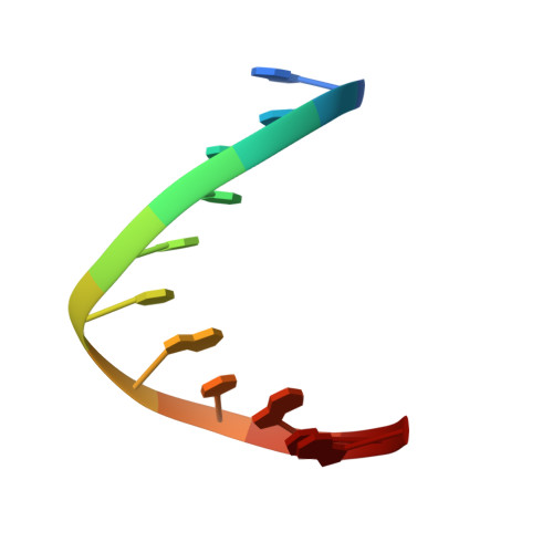
Lipid peroxidation products, as well as the metabolic products of vinyl chloride, react with cellular DNA producing the mutagenic adduct 3,N(4)-etheno-2'-deoxycytidine (epsilondC), along with several other exocyclic derivatives. High-resolution NMR spectroscopy and restrained molecular dynamics simulations were used to establish the solution structure of an 11-mer duplex containing an epsilondC.dC base-pair at its center. The NMR data suggested a regular right-handed helical structure having all residues in the anti orientation around the glycosydic torsion angle and Watson-Crick alignments for all canonical base-pairs of the duplex. Restrained molecular dynamics generated a three-dimensional model in excellent agreement with the spectroscopic data. The (epsilondC. dC)-duplex structure is a regular right-handed helix with a slight bend at the lesion site and no severe distortions of the sugar-phosphate backbone. The epsilondC adduct and its partner dC were displaced towards opposite grooves of the helix, resulting in a lesion-containing base-pair that was highly sheared but stabilized to some degree by the formation of a single hydrogen bond. Such a sheared base-pair alignment at the lesion site was previously observed for epsilondC.dG and epsilondC.T duplexes, and was also present in the crystal structures of duplexes containing dG.T and dG. U mismatches. These observations suggest the existence of a substrate structural motif that may be recognized by specific DNA glycosylases during the process of base excision repair.
Organizational Affiliation:
Department of Pharmacological Sciences, State University of New York at Stony Brook, Stony Brook, NY, 11794-8651, USA.