1B4F

OLIGOMERIC STRUCTURE OF THE HUMAN EPHB2 RECEPTOR SAM DOMAIN


Experimental Data Snapshot

  • Method: X-RAY DIFFRACTION
  • Resolution: 1.95 Å
  • R-Value Free: 0.273 
  • R-Value Work: 0.228 
  • R-Value Observed: 0.228 

wwPDB Validation   3D Report Full Report


This is version 1.4 of the entry. See complete history


Literature

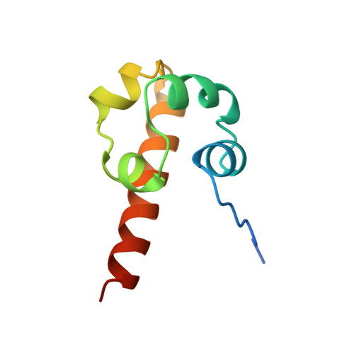
Oligomeric structure of the human EphB2 receptor SAM domain.

Thanos, C.D.Goodwill, K.E.Bowie, J.U.

(1999) Science 283: 833-836

  • DOI: https://doi.org/10.1126/science.283.5403.833
  • Primary Citation of Related Structures:  
    1B4F

  • PubMed Abstract: 

    The sterile alpha motif (SAM) domain is a protein interaction module that is present in diverse signal-transducing proteins. SAM domains are known to form homo- and hetero-oligomers. The crystal structure of the SAM domain from an Eph receptor tyrosine kinase, EphB2, reveals two large interfaces. In one interface, adjacent monomers exchange amino-terminal peptides that insert into a hydrophobic groove on each neighbor. A second interface is composed of the carboxyl-terminal helix and a nearby loop. A possible oligomer, constructed from a combination of these binding modes, may provide a platform for the formation of larger protein complexes.


  • Organizational Affiliation

    UCLA-DOE Laboratory of Structural Biology and Molecular Medicine and Department of Chemistry and Biochemistry, University of California, Los Angeles, CA 90095, USA.


Macromolecules
Find similar proteins by:  (by identity cutoff)  |  3D Structure
Entity ID: 1
MoleculeChains Sequence LengthOrganismDetailsImage
EPHB2
A, B, C, D, E
A, B, C, D, E, F, G, H
82Homo sapiensMutation(s): 0 
UniProt & NIH Common Fund Data Resources
Find proteins for P29323 (Homo sapiens)
Explore P29323 
Go to UniProtKB:  P29323
PHAROS:  P29323
GTEx:  ENSG00000133216 
Entity Groups  
Sequence Clusters30% Identity50% Identity70% Identity90% Identity95% Identity100% Identity
UniProt GroupP29323
Sequence Annotations
Expand
  • Reference Sequence
Experimental Data & Validation

Experimental Data

  • Method: X-RAY DIFFRACTION
  • Resolution: 1.95 Å
  • R-Value Free: 0.273 
  • R-Value Work: 0.228 
  • R-Value Observed: 0.228 
  • Space Group: P 41
Unit Cell:
Length ( Å )Angle ( ˚ )
a = 73.897α = 90
b = 73.897β = 90
c = 104.547γ = 90
Software Package:
Software NamePurpose
CNSrefinement
SHAKE-N-BAKEmodel building
DENZOdata reduction
SCALEPACKdata scaling
CNSphasing
SHAKE-N-BAKEphasing

Structure Validation

View Full Validation Report


Entry History 

Deposition Data

Revision History  (Full details and data files)

  • Version 1.0: 1999-02-16
    Type: Initial release
  • Version 1.1: 2008-03-24
    Changes: Version format compliance
  • Version 1.2: 2011-07-13
    Changes: Version format compliance
  • Version 1.3: 2019-12-11
    Changes: Advisory, Database references, Derived calculations
  • Version 1.4: 2024-02-07
    Changes: Data collection, Database references