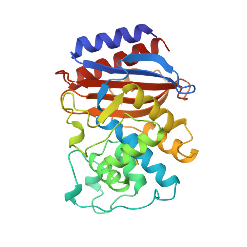
Crystal structure of an acylation transition-state analog of the TEM-1 beta-lactamase. Mechanistic implications for class A beta-lactamases.
Maveyraud, L., Pratt, R.F., Samama, J.P.(1998) Biochemistry 37: 2622-2628
- PubMed: 9485412
- DOI: https://doi.org/10.1021/bi972501b
- Primary Citation of Related Structures:
1AXB - PubMed Abstract:
The crystal structure of a phosphonate complex of the class A TEM-1 beta-lactamase has been determined to a resolution of 2.0 A. The phosphonate appears stoichiometrically at the active site, bound covalently to Ser70Ogamma, with one phosphonyl oxygen in the oxyanion hole. Although the overall structure is very similar to that of the native enzyme (rms difference 0.37 A for all heavy atoms), changes have occurred in the position of active site functional groups. The active site is also not in the conformation observed in the complex of another class A beta-lactamase, that of Staphylococcus aureus PC1, with the same phosphonate [Chen, C. C. H., et al. (1993) J. Mol. Biol. 234,165-178]. Both phosphonate structures, however, can be seen to represent models of acylation transition-states since in each the deacylating water molecule appears firmly bound to the Glu166 carboxylate group. The major difference between the structures lies in the positioning of Lys73Nzeta and Ser130Ogamma. In the S. aureus structure, the closest interaction of these functional groups is between Lys73Nzeta and Ser70Ogamma (2.8 A), while in the TEM-1 structure it is between Ser130Ogamma and the second phosphonyl oxygen of the bound inhibitor (2.8 A). The former structure therefore may resemble a transition state for formation of the tetrahedral species in acylation by nucleophilic attack on the substrate, where Lys73Nzeta presumably catalyzes the reaction as a general base. The TEM-1 structure can then be seen as an analogue of the transition state for breakdown of the tetrahedral species, where Ser130Ogamma is acting as a general acid, assisting the departure of the leaving group. The class A beta-lactamase crystal structures now available lead to a self-consistent proposal for a mechanism of catalysis by these enzymes.
Organizational Affiliation:
Groupe de Cristallographie Biologique, Institut de Pharmacologie et de Biologie Structurale, UPR 9062 CNRS, 205 route de Narbonne, 31077 Toulouse, France.