1AVO

PROTEASOME ACTIVATOR REG(ALPHA)


Experimental Data Snapshot

  • Method: X-RAY DIFFRACTION
  • Resolution: 2.80 Å
  • R-Value Free: 0.289 
  • R-Value Work: 0.249 
  • R-Value Observed: 0.249 

wwPDB Validation   3D Report Full Report


This is version 1.3 of the entry. See complete history


Literature

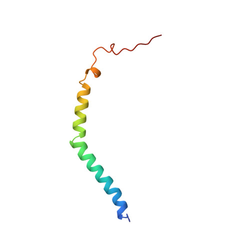
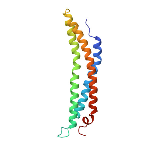
Structure of the proteasome activator REGalpha (PA28alpha).

Knowlton, J.R.Johnston, S.C.Whitby, F.G.Realini, C.Zhang, Z.Rechsteiner, M.Hill, C.P.

(1997) Nature 390: 639-643

  • DOI: https://doi.org/10.1038/37670
  • Primary Citation of Related Structures:  
    1AVO

  • PubMed Abstract: 

    The specificity of the 20S proteasome, which degrades many intracellular proteins, is regulated by protein complexes that bind to one or both ends of the cylindrical proteasome structure. One of these regulatory complexes, the 11S regulator (known as REG or PA28), stimulates proteasome peptidase activity and enhances the production of antigenic peptides for presentation by class I molecules of the major histocompatibility complex (MHC). The three REG subunits that have been identified, REGalpha, REGbeta and REGgamma (also known as the Ki antigen), share extensive sequence similarity, apart from a highly variable internal segment of 17-34 residues which may confer subunit-specific properties. REGalpha and REGbeta preferentially form a heteromeric complex, although purified REGalpha forms a heptamer in solution and has biochemical properties similar to the heteromeric REGalpha/REGbeta complex. We have now determined the crystal structure of human recombinant REGalpha at 2.8 A resolution. The heptameric barrel-shaped assembly contains a central channel that has an opening of 20 A diameter at one end and another of 30 A diameter at the presumed proteasome-binding surface. The binding of REG probably causes conformational changes that open a pore in the proteasome alpha-subunits through which substrates and products can pass.


  • Organizational Affiliation

    Department of Biochemistry, University of Utah, Salt Lake City 84132, USA.


Macromolecules
Find similar proteins by:  (by identity cutoff)  |  3D Structure
Entity ID: 1
MoleculeChains Sequence LengthOrganismDetailsImage
11S REGULATOR
A, C, E, G, I
A, C, E, G, I, K, M
60Homo sapiensMutation(s): 0 
UniProt & NIH Common Fund Data Resources
Find proteins for Q06323 (Homo sapiens)
Explore Q06323 
Go to UniProtKB:  Q06323
PHAROS:  Q06323
GTEx:  ENSG00000092010 
Entity Groups  
Sequence Clusters30% Identity50% Identity70% Identity90% Identity95% Identity100% Identity
UniProt GroupQ06323
Sequence Annotations
Expand
  • Reference Sequence
Find similar proteins by:  (by identity cutoff)  |  3D Structure
Entity ID: 2
MoleculeChains Sequence LengthOrganismDetailsImage
11S REGULATOR
B, D, F, H, J
B, D, F, H, J, L, N
140Homo sapiensMutation(s): 0 
UniProt & NIH Common Fund Data Resources
Find proteins for Q06323 (Homo sapiens)
Explore Q06323 
Go to UniProtKB:  Q06323
PHAROS:  Q06323
GTEx:  ENSG00000092010 
Entity Groups  
Sequence Clusters30% Identity50% Identity70% Identity90% Identity95% Identity100% Identity
UniProt GroupQ06323
Sequence Annotations
Expand
  • Reference Sequence
Experimental Data & Validation

Experimental Data

  • Method: X-RAY DIFFRACTION
  • Resolution: 2.80 Å
  • R-Value Free: 0.289 
  • R-Value Work: 0.249 
  • R-Value Observed: 0.249 
  • Space Group: C 1 2 1
Unit Cell:
Length ( Å )Angle ( ˚ )
a = 162.6α = 90
b = 134.3β = 90.6
c = 116.2γ = 90
Software Package:
Software NamePurpose
X-PLORmodel building
X-PLORrefinement
DENZOdata reduction
SCALEPACKdata scaling
X-PLORphasing

Structure Validation

View Full Validation Report


Entry History 

Deposition Data

Revision History  (Full details and data files)

  • Version 1.0: 1997-12-31
    Type: Initial release
  • Version 1.1: 2008-03-24
    Changes: Version format compliance
  • Version 1.2: 2011-07-13
    Changes: Version format compliance
  • Version 1.3: 2024-02-07
    Changes: Data collection, Database references, Other