1ART

X-RAY CRYSTALLOGRAPHIC STUDY OF PYRIDOXAL 5'-PHOSPHATE-TYPE ASPARTATE AMINOTRANSFERASES FROM ESCHERICHIA COLI IN OPEN AND CLOSED FORM


Experimental Data Snapshot

  • Method: X-RAY DIFFRACTION
  • Resolution: 1.80 Å
  • R-Value Work: 0.217 
  • R-Value Observed: 0.217 

wwPDB Validation   3D Report Full Report


This is version 1.4 of the entry. See complete history


Literature

X-ray crystallographic study of pyridoxal 5'-phosphate-type aspartate aminotransferases from Escherichia coli in open and closed form.

Okamoto, A.Higuchi, T.Hirotsu, K.Kuramitsu, S.Kagamiyama, H.

(1994) J Biochem 116: 95-107

  • DOI: https://doi.org/10.1093/oxfordjournals.jbchem.a124509
  • Primary Citation of Related Structures:  
    1ARS, 1ART

  • PubMed Abstract: 

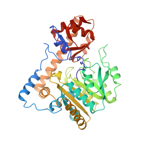
    We determined the three-dimensional structures of aspartate aminotransferase (AspAT) from Escherichia coli and its complex with inhibitor (2-methyl-L-aspartate) at 1.8A resolution. This enzyme reversibly catalyzes the transamination reaction and is a dimer of two identical subunits. Each subunit has 396 amino acid residues and one pyridoxal 5'-phosphate as a cofactor, and is divided into two domains, one large and the other small. Upon binding of the inhibitor, the small domain rotates by 5 degrees toward the large domain to close the active site. This domain movement is caused mainly by small but important main-chain conformational changes in the residues located over the domain interface of the small domain. In chicken mitochondrial AspAT, the domain movement was larger, with a rotational angle of 13 degrees. By comparison of these two structures, the difference in the rotational angles was found to be caused by the larger opening of the domain in the open form of chicken mitochondrial AspAT. Although the overall structures of these two enzymes were almost identical, the surface area of the domain interface in the E. coli enzyme was larger than that in mitochondrial AspAT, suggesting that the structure of the domain interface is responsible for the degree of movement of the small domain.


  • Organizational Affiliation

    Department of Chemistry, Faculty of Science, Osaka City University.


Macromolecules
Find similar proteins by:  (by identity cutoff)  |  3D Structure
Entity ID: 1
MoleculeChains Sequence LengthOrganismDetailsImage
ASPARTATE AMINOTRANSFERASE396Escherichia coliMutation(s): 0 
EC: 2.6.1.1
UniProt
Find proteins for P00509 (Escherichia coli (strain K12))
Explore P00509 
Go to UniProtKB:  P00509
Entity Groups  
Sequence Clusters30% Identity50% Identity70% Identity90% Identity95% Identity100% Identity
UniProt GroupP00509
Sequence Annotations
Expand
  • Reference Sequence
Small Molecules
Ligands 2 Unique
IDChains Name / Formula / InChI Key2D Diagram3D Interactions
PLP
Query on PLP

Download Ideal Coordinates CCD File 
B [auth A]PYRIDOXAL-5'-PHOSPHATE
C8 H10 N O6 P
NGVDGCNFYWLIFO-UHFFFAOYSA-N
0A0
Query on 0A0

Download Ideal Coordinates CCD File 
C [auth A]2-methyl-L-aspartic acid
C5 H9 N O4
CWAYDJFPMMUKOI-YFKPBYRVSA-N
Experimental Data & Validation

Experimental Data

  • Method: X-RAY DIFFRACTION
  • Resolution: 1.80 Å
  • R-Value Work: 0.217 
  • R-Value Observed: 0.217 
  • Space Group: C 2 2 21
Unit Cell:
Length ( Å )Angle ( ˚ )
a = 157.14α = 90
b = 85.51β = 90
c = 78.93γ = 90
Software Package:
Software NamePurpose
X-PLORmodel building
X-PLORrefinement
X-PLORphasing

Structure Validation

View Full Validation Report


Entry History 

Deposition Data

Revision History  (Full details and data files)

  • Version 1.0: 1994-08-31
    Type: Initial release
  • Version 1.1: 2008-03-03
    Changes: Version format compliance
  • Version 1.2: 2011-07-13
    Changes: Derived calculations, Version format compliance
  • Version 1.3: 2017-11-29
    Changes: Derived calculations, Other
  • Version 1.4: 2024-02-07
    Changes: Data collection, Database references, Derived calculations