1ANX

THE CRYSTAL STRUCTURE OF A NEW HIGH-CALCIUM FORM OF ANNEXIN V


Experimental Data Snapshot

  • Method: X-RAY DIFFRACTION
  • Resolution: 1.90 Å
  • R-Value Observed: 0.198 

wwPDB Validation   3D Report Full Report


This is version 1.3 of the entry. See complete history


Literature

The crystal structure of a new high-calcium form of annexin V.

Sopkova, J.Renouard, M.Lewit-Bentley, A.

(1993) J Mol Biol 234: 816-825

  • DOI: https://doi.org/10.1006/jmbi.1993.1627
  • Primary Citation of Related Structures:  
    1ANX

  • PubMed Abstract: 

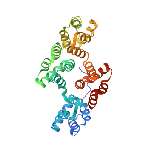
    Annexin V was crystallized in the presence of a high concentration of calcium and the structure refined at 1.9 A resolution. The crystals are triclinic (P1) with three molecules per asymmetric unit and pseudo-R3 symmetry, reflecting a tendency of annexin to form trimers. The overall structure of the protein is similar to that seen in other crystal forms. There are, however, significant changes in domain III, where a new calcium site is formed. The whole region surrounding this site is reorganized in our structure, rendering annexin V more symmetrical and more alike annexin I. The formation of the new calcium site causes the displacement of Trp187 from a buried to an exposed conformation, a change that has recently been demonstrated by fluorescence measurements. The affinity of the different potential calcium sites is modulated: there is no calcium bound in domains II and IV, while up to two secondary calcium ions sites (in domains I and III) can substitute, depending on the calcium concentration present. We suggest that annexin can act as a calcium buffer, binding or releasing calcium depending on its local concentration. Our results also show that annexin displays inherent mobility which, together with its capacity to modulate the calcium affinity of its sites, can be of importance for its function on the membrane surface.


  • Organizational Affiliation

    LURE, Centre Universitaire Paris-Sud, Orsay, France.


Macromolecules
Find similar proteins by:  (by identity cutoff)  |  3D Structure
Entity ID: 1
MoleculeChains Sequence LengthOrganismDetailsImage
ANNEXIN V
A, B, C
319Homo sapiensMutation(s): 0 
UniProt & NIH Common Fund Data Resources
Find proteins for P08758 (Homo sapiens)
Explore P08758 
Go to UniProtKB:  P08758
PHAROS:  P08758
GTEx:  ENSG00000164111 
Entity Groups  
Sequence Clusters30% Identity50% Identity70% Identity90% Identity95% Identity100% Identity
UniProt GroupP08758
Sequence Annotations
Expand
  • Reference Sequence
Small Molecules
Ligands 2 Unique
IDChains Name / Formula / InChI Key2D Diagram3D Interactions
SO4
Query on SO4

Download Ideal Coordinates CCD File 
E [auth A],
J [auth B],
O [auth C]
SULFATE ION
O4 S
QAOWNCQODCNURD-UHFFFAOYSA-L
CA
Query on CA

Download Ideal Coordinates CCD File 
D [auth A]
F [auth A]
G [auth A]
H [auth A]
I [auth B]
D [auth A],
F [auth A],
G [auth A],
H [auth A],
I [auth B],
K [auth B],
L [auth B],
M [auth B],
N [auth C],
P [auth C],
Q [auth C],
R [auth C]
CALCIUM ION
Ca
BHPQYMZQTOCNFJ-UHFFFAOYSA-N
Experimental Data & Validation

Experimental Data

  • Method: X-RAY DIFFRACTION
  • Resolution: 1.90 Å
  • R-Value Observed: 0.198 
  • Space Group: P 1
Unit Cell:
Length ( Å )Angle ( ˚ )
a = 91.2α = 82.4
b = 91.3β = 82.4
c = 36.2γ = 118.2
Software Package:
Software NamePurpose
PROLSQrefinement

Structure Validation

View Full Validation Report


Entry History 

Deposition Data

Revision History  (Full details and data files)

  • Version 1.0: 1994-12-20
    Type: Initial release
  • Version 1.1: 2008-03-24
    Changes: Version format compliance
  • Version 1.2: 2011-07-13
    Changes: Version format compliance
  • Version 1.3: 2024-02-07
    Changes: Data collection, Database references, Derived calculations, Other