1AI8

HUMAN ALPHA-THROMBIN TERNARY COMPLEX WITH THE EXOSITE INHIBITOR HIRUGEN AND ACTIVE SITE INHIBITOR PHCH2OCO-D-DPA-PRO-BOROMPG


Experimental Data Snapshot

  • Method: X-RAY DIFFRACTION
  • Resolution: 1.85 Å
  • R-Value Free: 0.240 
  • R-Value Work: 0.170 

wwPDB Validation   3D Report Full Report


This is version 1.3 of the entry. See complete history


Literature

The refined 1.9-A X-ray crystal structure of D-Phe-Pro-Arg chloromethylketone-inhibited human alpha-thrombin: structure analysis, overall structure, electrostatic properties, detailed active-site geometry, and structure-function relationships.

Bode, W.Turk, D.Karshikov, A.

(1992) Protein Sci 1: 426-471

  • DOI: https://doi.org/10.1002/pro.5560010402
  • Primary Citation of Related Structures:  
    1AI8, 1AIX

  • PubMed Abstract: 

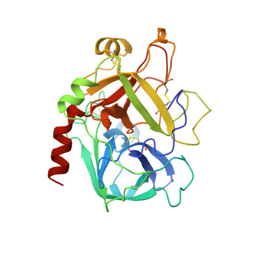
    Thrombin is a multifunctional serine proteinase that plays a key role in coagulation while exhibiting several other key cellular bioregulatory functions. The X-ray crystal structure of human alpha-thrombin was determined in its complex with the specific thrombin inhibitor D-Phe-Pro-Arg chloromethylketone (PPACK) using Patterson search methods and a search model derived from trypsinlike proteinases of known spatial structure (Bode, W., Mayr, I., Baumann, U., Huber, R., Stone, S.R., & Hofsteenge, J., 1989, EMBO J. 8, 3467-3475). The crystallographic refinement of the PPACK-thrombin model has now been completed at an R value of 0.156 (8 to 1.92 A); in particular, the amino- and the carboxy-termini of the thrombin A-chain are now defined and all side-chain atoms localized; only proline 37 was found to be in a cis-peptidyl conformation. The thrombin B-chain exhibits the characteristic polypeptide fold of trypsinlike serine proteinases; 195 residues occupy topologically equivalent positions with residues in bovine trypsin and 190 with those in bovine chymotrypsin with a root-mean-square (r.m.s.) deviation of 0.8 A for their alpha-carbon atoms. Most of the inserted residues constitute novel surface loops. A chymotrypsinogen numbering is suggested for thrombin based on the topological equivalences. The thrombin A-chain is arranged in a boomeranglike shape against the B-chain globule opposite to the active site; it resembles somewhat the propeptide of chymotrypsin(ogen) and is similarly not involved in substrate and inhibitor binding. Thrombin possesses an exceptionally large proportion of charged residues. The negatively and positively charged residues are not distributed uniformly over the whole molecule, but are clustered to form a sandwichlike electrostatic potential; in particular, two extended patches of mainly positively charged residues occur close to the carboxy-terminal B-chain helix (forming the presumed heparin-binding site) and on the surface of loop segment 70-80 (the fibrin[ogen] secondary binding exosite), respectively; the negatively charged residues are more clustered in the ringlike region between both poles, particularly around the active site. Several of the charged residues are involved in salt bridges; most are on the surface, but 10 charged protein groups form completely buried salt bridges and clusters. These electrostatic interactions play a particularly important role in the intrachain stabilization of the A-chain, in the coherence between the A- and the B-chain, and in the surface structure of the fibrin(ogen) secondary binding exosite (loop segment 67-80).(ABSTRACT TRUNCATED AT 400 WORDS)


  • Organizational Affiliation

    Max-Planck-Institut für Biochemie, Martinsried, Germany.


Macromolecules
Find similar proteins by:  (by identity cutoff)  |  3D Structure
Entity ID: 1
MoleculeChains Sequence LengthOrganismDetailsImage
ALPHA-THROMBIN (SMALL SUBUNIT)A [auth L]36Homo sapiensMutation(s): 0 
EC: 3.4.21.5
UniProt & NIH Common Fund Data Resources
Find proteins for P00734 (Homo sapiens)
Explore P00734 
Go to UniProtKB:  P00734
PHAROS:  P00734
GTEx:  ENSG00000180210 
Entity Groups  
Sequence Clusters30% Identity50% Identity70% Identity90% Identity95% Identity100% Identity
UniProt GroupP00734
Sequence Annotations
Expand
  • Reference Sequence
Find similar proteins by:  (by identity cutoff)  |  3D Structure
Entity ID: 2
MoleculeChains Sequence LengthOrganismDetailsImage
ALPHA-THROMBIN (LARGE SUBUNIT)B [auth H]259Homo sapiensMutation(s): 0 
EC: 3.4.21.5
UniProt & NIH Common Fund Data Resources
Find proteins for P00734 (Homo sapiens)
Explore P00734 
Go to UniProtKB:  P00734
PHAROS:  P00734
GTEx:  ENSG00000180210 
Entity Groups  
Sequence Clusters30% Identity50% Identity70% Identity90% Identity95% Identity100% Identity
UniProt GroupP00734
Sequence Annotations
Expand
  • Reference Sequence

Find similar proteins by:  Sequence   |   3D Structure  

Entity ID: 3
MoleculeChains Sequence LengthOrganismDetailsImage
HIRUDIN IIIBC [auth I]12Hirudo medicinalisMutation(s): 0 
EC: 3.4.21.5
UniProt
Find proteins for P01050 (Hirudo medicinalis)
Explore P01050 
Go to UniProtKB:  P01050
Entity Groups  
Sequence Clusters30% Identity50% Identity70% Identity90% Identity95% Identity100% Identity
UniProt GroupP01050
Sequence Annotations
Expand
  • Reference Sequence
Small Molecules
Ligands 1 Unique
IDChains Name / Formula / InChI Key2D Diagram3D Interactions
T42
Query on T42

Download Ideal Coordinates CCD File 
D [auth H]MORPHOLINO-DIPHENYLALANINE-METHOXYPROPYLBORONIC ACID
C30 H39 B N4 O7
DTJORIMAIDQZOF-NSROOHRNSA-N
Modified Residues  1 Unique
IDChains TypeFormula2D DiagramParent
TYS
Query on TYS
C [auth I]L-PEPTIDE LINKINGC9 H11 N O6 STYR
Experimental Data & Validation

Experimental Data

  • Method: X-RAY DIFFRACTION
  • Resolution: 1.85 Å
  • R-Value Free: 0.240 
  • R-Value Work: 0.170 
  • Space Group: C 1 2 1
Unit Cell:
Length ( Å )Angle ( ˚ )
a = 69.714α = 90
b = 71.464β = 99.82
c = 71.648γ = 90
Software Package:
Software NamePurpose
DENZOdata reduction
SCALEPACKdata scaling
AMoREphasing
REFMACrefinement

Structure Validation

View Full Validation Report


Entry History 

Deposition Data

Revision History  (Full details and data files)

  • Version 1.0: 1997-10-15
    Type: Initial release
  • Version 1.1: 2008-03-03
    Changes: Version format compliance
  • Version 1.2: 2011-07-13
    Changes: Atomic model, Database references, Derived calculations, Non-polymer description, Structure summary, Version format compliance
  • Version 1.3: 2023-08-02
    Changes: Database references, Derived calculations, Refinement description