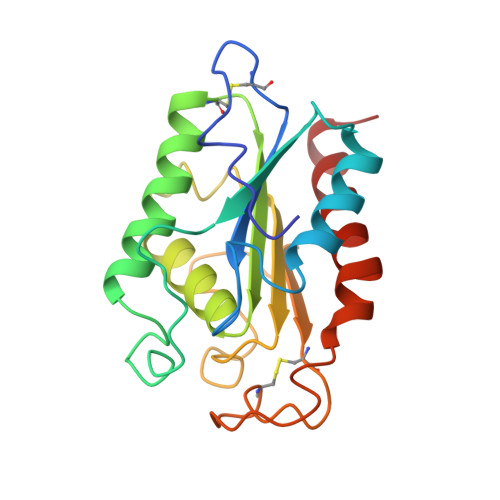
Atomic resolution (1.0 A) crystal structure of Fusarium solani cutinase: stereochemical analysis.
Longhi, S., Czjzek, M., Lamzin, V., Nicolas, A., Cambillau, C.(1997) J Mol Biol 268: 779-799
- PubMed: 9175860
- DOI: https://doi.org/10.1006/jmbi.1997.1000
- Primary Citation of Related Structures:
1AGY, 1CEX - PubMed Abstract:
X-ray data have been recorded to 1.0 A resolution from a crystal of Fusarium solani cutinase using synchrotron radiation and an imaging-plate scanner. The anisotropic treatment of thermal motion led to a fivefold increase in accuracy and to a considerable quality improvement in the electron density maps with respect to an intermediate isotropic model. The final model has an R-factor of 9.4%, with a mean coordinate error of 0.021 A, as estimated from inversion of the least-squares matrix. The availability of an accurate structure at atomic resolution and of meaningful estimates of the errors in its atomic parameters, allowed an extensive analysis of several stereochemical parameters, such as peptide planarity, main-chain and some side-chain bond distances. The hydrogen atoms could be clearly identified in the electron density, thus providing unambiguous evidence on the protonation state of the catalytic histidine residue. The atomic resolution revealed an appreciable extent of flexibility in the cutinase active site, which might be correlated with a possible adaptation to different substrates. The anisotropic treatment of thermal factors provided insights into the anisotropic nature of motions. The analysis of these motions in the two loops delimiting the catalytic crevice pointed out a "breath-like" movement in the substrate binding region of cutinase.
Organizational Affiliation:
Architecture et Fonction des Macromolécules Biologiques UPR 9039, AFMB-CNRS, IFR1, Marseille, France.