1ZXN

Human DNA topoisomerase IIa ATPase/ADP


Experimental Data Snapshot

  • Method: X-RAY DIFFRACTION
  • Resolution: 2.51 Å
  • R-Value Free: 0.281 
  • R-Value Work: 0.235 
  • R-Value Observed: 0.235 

wwPDB Validation   3D Report Full Report


Ligand Structure Quality Assessment 


This is version 1.3 of the entry. See complete history

Re-refinement Note

A newer entry is available that reflects an alternative modeling of the original data: 4R1F


Literature

Nucleotide-dependent Domain Movement in the ATPase Domain of a Human Type IIA DNA Topoisomerase.

Wei, H.Ruthenburg, A.J.Bechis, S.K.Verdine, G.L.

(2005) J Biol Chem 280: 37041-37047

  • DOI: https://doi.org/10.1074/jbc.M506520200
  • Primary Citation of Related Structures:  
    1ZXM, 1ZXN

  • PubMed Abstract: 

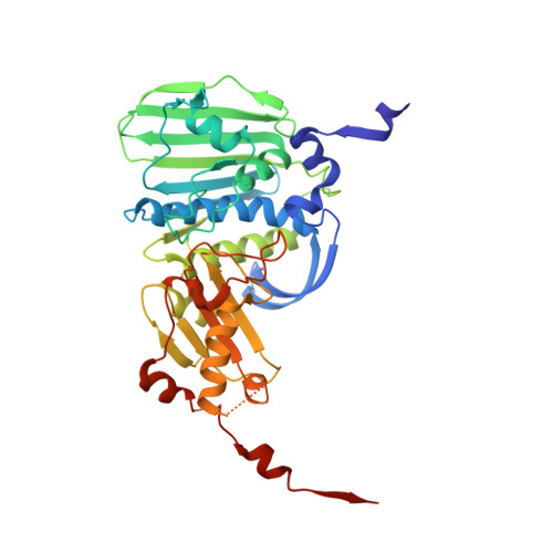
    Type IIA DNA topoisomerases play multiple essential roles in the management of higher-order DNA structure, including modulation of topological state, chromosome segregation, and chromatin condensation. These diverse physiologic functions are all accomplished through a common molecular mechanism, wherein the protein catalyzes transient cleavage of a DNA duplex (the G-segment) to yield a double-stranded gap through which another duplex (the T-segment) is passed. The overall process is orchestrated by the opening and closing of molecular "gates" in the topoisomerase structure, which is regulated by ATP binding, hydrolysis, and release of ADP and inorganic phosphate. Here we present two crystal structures of the ATPase domain of human DNA topoisomerase IIalpha in different nucleotide-bound states. Comparison of these structures revealed rigid-body movement of the structural modules within the ATPase domain, suggestive of the motions of a molecular gate.


  • Organizational Affiliation

    Department of Chemistry and Chemical Biology, Harvard University, Cambridge, Massachusetts 02138, USA.


Macromolecules
Find similar proteins by:  (by identity cutoff)  |  3D Structure
Entity ID: 1
MoleculeChains Sequence LengthOrganismDetailsImage
DNA topoisomerase II, alpha isozyme
A, B, C, D
400Homo sapiensMutation(s): 0 
Gene Names: TOP2ATOP2
EC: 5.99.1.3
UniProt & NIH Common Fund Data Resources
Find proteins for P11388 (Homo sapiens)
Explore P11388 
Go to UniProtKB:  P11388
PHAROS:  P11388
GTEx:  ENSG00000131747 
Entity Groups  
Sequence Clusters30% Identity50% Identity70% Identity90% Identity95% Identity100% Identity
UniProt GroupP11388
Sequence Annotations
Expand
  • Reference Sequence
Small Molecules
Ligands 4 Unique
IDChains Name / Formula / InChI Key2D Diagram3D Interactions
ADP
Query on ADP

Download Ideal Coordinates CCD File 
F [auth A],
J [auth B],
L [auth C],
O [auth D]
ADENOSINE-5'-DIPHOSPHATE
C10 H15 N5 O10 P2
XTWYTFMLZFPYCI-KQYNXXCUSA-N
SO4
Query on SO4

Download Ideal Coordinates CCD File 
I [auth B],
N [auth D]
SULFATE ION
O4 S
QAOWNCQODCNURD-UHFFFAOYSA-L
GOL
Query on GOL

Download Ideal Coordinates CCD File 
G [auth A]GLYCEROL
C3 H8 O3
PEDCQBHIVMGVHV-UHFFFAOYSA-N
MG
Query on MG

Download Ideal Coordinates CCD File 
E [auth A],
H [auth B],
K [auth C],
M [auth D]
MAGNESIUM ION
Mg
JLVVSXFLKOJNIY-UHFFFAOYSA-N
Experimental Data & Validation

Experimental Data

  • Method: X-RAY DIFFRACTION
  • Resolution: 2.51 Å
  • R-Value Free: 0.281 
  • R-Value Work: 0.235 
  • R-Value Observed: 0.235 
  • Space Group: P 1 21 1
Unit Cell:
Length ( Å )Angle ( ˚ )
a = 71.015α = 90
b = 90.492β = 89.97
c = 148.293γ = 90
Software Package:
Software NamePurpose
HKL-2000data collection
SCALEPACKdata scaling
AMoREphasing
CNSrefinement
HKL-2000data reduction

Structure Validation

View Full Validation Report


Ligand Structure Quality Assessment 


Entry History 

Deposition Data

Revision History  (Full details and data files)

  • Version 1.0: 2005-08-23
    Type: Initial release
  • Version 1.1: 2008-04-30
    Changes: Version format compliance
  • Version 1.2: 2011-07-13
    Changes: Non-polymer description, Version format compliance
  • Version 1.3: 2024-02-14
    Changes: Advisory, Data collection, Database references, Derived calculations