1ZT7

crystal structure of class I MHC H-2Kk in complex with a nonapeptide


Experimental Data Snapshot

  • Method: X-RAY DIFFRACTION
  • Resolution: 3.00 Å
  • R-Value Free: 0.311 
  • R-Value Work: 0.231 
  • R-Value Observed: 0.235 

wwPDB Validation   3D Report Full Report


This is version 1.4 of the entry. See complete history


Literature

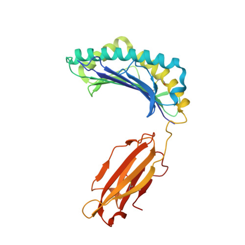
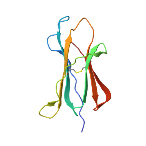
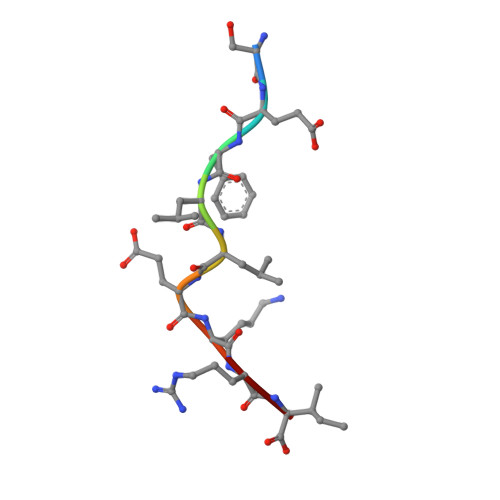
The H-2Kk MHC peptide-binding groove anchors the backbone of an octameric antigenic peptide in an unprecedented mode.

Kellenberger, C.Roussel, A.Malissen, B.

(2005) J Immunol 175: 3819-3825

  • DOI: https://doi.org/10.4049/jimmunol.175.6.3819
  • Primary Citation of Related Structures:  
    1ZT1, 1ZT7

  • PubMed Abstract: 

    A wealth of data has accumulated on the structure of mouse MHC class I (MHCI) molecules encoded by the H-2(b) and H-2(d) haplotypes. In contrast, there is a dearth of structural data regarding H-2(k)-encoded molecules. Therefore, the structures of H-2K(k) complexed to an octameric peptide from influenza A virus (HA(259-266)) and to a nonameric peptide from SV40 (SV40(560-568)) have been determined by x-ray crystallography at 2.5 and 3.0 A resolutions, respectively. The structure of the H-2K(k)-HA(259-266) complex reveals that residues located on the floor of the peptide-binding groove contact directly the backbone of the octameric peptide and force it to lie deep within the H-2K(k) groove. This unprecedented mode of peptide binding occurs despite the presence of bulky residues in the middle of the floor of the H-2K(k) peptide-binding groove. As a result, the Calpha atoms of peptide residues P5 and P6 are more buried than the corresponding residues of H-2K(b)-bound octapeptides, making them even less accessible to TCR contact. When bound to H-2K(k), the backbone of the SV40(560-568) nonapeptide bulges out of the peptide-binding groove and adopts a conformation reminiscent of that observed for peptides bound to H-2L(d). This structural convergence occurs despite the totally different architectures of the H-2L(d) and H-2K(k) peptide-binding grooves. Therefore, these two H-2K(k)-peptide complexes provide insights into the mechanisms through which MHC polymorphism outside primary peptide pockets influences the conformation of the bound peptides and have implications for TCR recognition and vaccine design.


  • Organizational Affiliation

    Centre d'Immunologie de Marseille-Luminy, Institut National de la Santé et de la Recherche Médicale-Centre National Recherche de la Scientifique-Université de la Méditerranée, Parc Scientifique de Luminy, Marseille, France. kellenberger@ciml.univ-mrs.fr


Macromolecules
Find similar proteins by:  (by identity cutoff)  |  3D Structure
Entity ID: 1
MoleculeChains Sequence LengthOrganismDetailsImage
H-2 class I histocompatibility antigen, K-K alpha chain
A, C
276Mus musculusMutation(s): 0 
UniProt
Find proteins for P04223 (Mus musculus)
Explore P04223 
Go to UniProtKB:  P04223
Entity Groups  
Sequence Clusters30% Identity50% Identity70% Identity90% Identity95% Identity100% Identity
UniProt GroupP04223
Sequence Annotations
Expand
  • Reference Sequence
Find similar proteins by:  (by identity cutoff)  |  3D Structure
Entity ID: 2
MoleculeChains Sequence LengthOrganismDetailsImage
Beta-2-microglobulin
B, D
100Mus musculusMutation(s): 0 
UniProt & NIH Common Fund Data Resources
Find proteins for P01887 (Mus musculus)
Explore P01887 
Go to UniProtKB:  P01887
IMPC:  MGI:88127
Entity Groups  
Sequence Clusters30% Identity50% Identity70% Identity90% Identity95% Identity100% Identity
UniProt GroupP01887
Sequence Annotations
Expand
  • Reference Sequence

Find similar proteins by:  Sequence   |   3D Structure  

Entity ID: 3
MoleculeChains Sequence LengthOrganismDetailsImage
SV40 epitope, SEFLLEKRIE [auth P],
F [auth Q]
9N/AMutation(s): 0 
Sequence Annotations
Expand
  • Reference Sequence
Experimental Data & Validation

Experimental Data

  • Method: X-RAY DIFFRACTION
  • Resolution: 3.00 Å
  • R-Value Free: 0.311 
  • R-Value Work: 0.231 
  • R-Value Observed: 0.235 
  • Space Group: P 1 2 1
Unit Cell:
Length ( Å )Angle ( ˚ )
a = 85.135α = 90
b = 72.628β = 111.24
c = 88.793γ = 90
Software Package:
Software NamePurpose
REFMACrefinement
DENZOdata reduction
SCALEPACKdata scaling
AMoREphasing

Structure Validation

View Full Validation Report


Entry History 

Deposition Data

Revision History  (Full details and data files)

  • Version 1.0: 2005-10-18
    Type: Initial release
  • Version 1.1: 2008-04-30
    Changes: Version format compliance
  • Version 1.2: 2011-07-13
    Changes: Version format compliance
  • Version 1.3: 2019-11-06
    Changes: Data collection, Database references
  • Version 1.4: 2024-04-03
    Changes: Data collection, Database references, Refinement description