1ZS8

Crystal Structure of the Murine MHC Class Ib Molecule M10.5


Experimental Data Snapshot

  • Method: X-RAY DIFFRACTION
  • Resolution: 3.00 Å
  • R-Value Free: 0.308 
  • R-Value Work: 0.307 
  • R-Value Observed: 0.307 

wwPDB Validation   3D Report Full Report


Ligand Structure Quality Assessment 


This is version 1.5 of the entry. See complete history


Literature

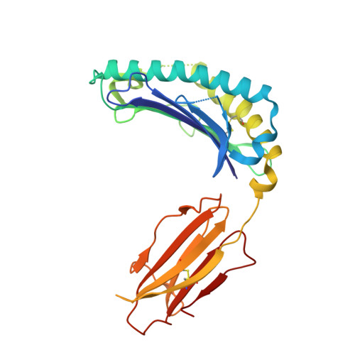
Structure of a pheromone receptor-associated MHC molecule with an open and empty groove.

Olson, R.Huey-Tubman, K.E.Dulac, C.Bjorkman, P.J.

(2005) PLoS Biol 3: e257-e257

  • DOI: https://doi.org/10.1371/journal.pbio.0030257
  • Primary Citation of Related Structures:  
    1ZS8

  • PubMed Abstract: 

    Neurons in the murine vomeronasal organ (VNO) express a family of class Ib major histocompatibility complex (MHC) proteins (M10s) that interact with the V2R class of VNO receptors. This interaction may play a direct role in the detection of pheromonal cues that initiate reproductive and territorial behaviors. The crystal structure of M10.5, an M10 family member, is similar to that of classical MHC molecules. However, the M10.5 counterpart of the MHC peptide-binding groove is open and unoccupied, revealing the first structure of an empty class I MHC molecule. Similar to empty MHC molecules, but unlike peptide-filled MHC proteins and non-peptide-binding MHC homologs, M10.5 is thermally unstable, suggesting that its groove is normally occupied. However, M10.5 does not bind endogenous peptides when expressed in mammalian cells or when offered a mixture of class I-binding peptides. The F pocket side of the M10.5 groove is open, suggesting that ligands larger than 8-10-mer class I-binding peptides could fit by extending out of the groove. Moreover, variable residues point up from the groove helices, rather than toward the groove as in classical MHC structures. These data suggest that M10s are unlikely to provide specific recognition of class I MHC-binding peptides, but are consistent with binding to other ligands, including proteins such as the V2Rs.


  • Organizational Affiliation

    Division of Biology, California Institute of Technology, Pasadena, California, USA.


Macromolecules
Find similar proteins by:  (by identity cutoff)  |  3D Structure
Entity ID: 1
MoleculeChains Sequence LengthOrganismDetailsImage
histocompatibility 2, M region locus 10.5
A, C, E, G, I
274Mus musculusMutation(s): 0 
Gene Names: M10.5
UniProt
Find proteins for Q860W6 (Mus musculus)
Explore Q860W6 
Go to UniProtKB:  Q860W6
Entity Groups  
Sequence Clusters30% Identity50% Identity70% Identity90% Identity95% Identity100% Identity
UniProt GroupQ860W6
Sequence Annotations
Expand
  • Reference Sequence
Find similar proteins by:  (by identity cutoff)  |  3D Structure
Entity ID: 2
MoleculeChains Sequence LengthOrganismDetailsImage
Beta-2-microglobulin
B, D, F, H, J
99Homo sapiensMutation(s): 0 
Gene Names: B2M
UniProt & NIH Common Fund Data Resources
Find proteins for P61769 (Homo sapiens)
Explore P61769 
Go to UniProtKB:  P61769
PHAROS:  P61769
GTEx:  ENSG00000166710 
Entity Groups  
Sequence Clusters30% Identity50% Identity70% Identity90% Identity95% Identity100% Identity
UniProt GroupP61769
Sequence Annotations
Expand
  • Reference Sequence
Experimental Data & Validation

Experimental Data

  • Method: X-RAY DIFFRACTION
  • Resolution: 3.00 Å
  • R-Value Free: 0.308 
  • R-Value Work: 0.307 
  • R-Value Observed: 0.307 
  • Space Group: P 21 21 21
Unit Cell:
Length ( Å )Angle ( ˚ )
a = 124.11α = 90
b = 134.71β = 90
c = 149.37γ = 90
Software Package:
Software NamePurpose
DENZOdata reduction
SCALEPACKdata scaling
AMoREphasing
CNSrefinement
PDB_EXTRACTdata extraction

Structure Validation

View Full Validation Report


Ligand Structure Quality Assessment 


Entry History 

Deposition Data

Revision History  (Full details and data files)

  • Version 1.0: 2005-07-26
    Type: Initial release
  • Version 1.1: 2008-04-30
    Changes: Version format compliance
  • Version 1.2: 2011-07-13
    Changes: Non-polymer description, Version format compliance
  • Version 1.3: 2017-10-11
    Changes: Refinement description
  • Version 1.4: 2020-07-29
    Type: Remediation
    Reason: Carbohydrate remediation
    Changes: Data collection, Derived calculations, Structure summary
  • Version 1.5: 2023-08-23
    Changes: Data collection, Database references, Refinement description, Structure summary