1ZP3

E. coli Methylenetetrahydrofolate Reductase (oxidized)


Experimental Data Snapshot

  • Method: X-RAY DIFFRACTION
  • Resolution: 1.85 Å
  • R-Value Free: 0.237 
  • R-Value Work: 0.214 
  • R-Value Observed: 0.214 

wwPDB Validation   3D Report Full Report


Ligand Structure Quality Assessment 


This is version 1.4 of the entry. See complete history


Literature

Structures of NADH and CH(3)-H(4)Folate Complexes of Escherichia coli Methylenetetrahydrofolate Reductase Reveal a Spartan Strategy for a Ping-Pong Reaction

Pejchal, R.Sargeant, R.Ludwig, M.L.

(2005) Biochemistry 44: 11447-11457

  • DOI: https://doi.org/10.1021/bi050533q
  • Primary Citation of Related Structures:  
    1ZP3, 1ZP4, 1ZPT, 1ZRQ

  • PubMed Abstract: 

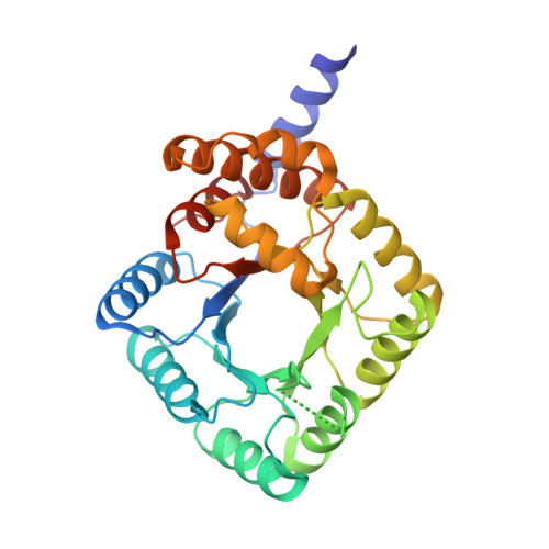
    Methylenetetrahydrofolate reductases (MTHFRs; EC 1.7.99.5) catalyze the NAD(P)H-dependent reduction of 5,10-methylenetetrahydrofolate (CH(2)-H(4)folate) to 5-methyltetrahydrofolate (CH(3)-H(4)folate) using flavin adenine dinucleotide (FAD) as a cofactor. The initial X-ray structure of Escherichia coli MTHFR revealed that this 33-kDa polypeptide is a (betaalpha)(8) barrel that aggregates to form an unusual tetramer with only 2-fold symmetry. Structures of reduced enzyme complexed with NADH and of oxidized Glu28Gln enzyme complexed with CH(3)-H(4)folate have now been determined at resolutions of 1.95 and 1.85 A, respectively. The NADH complex reveals a rare mode of dinucleotide binding; NADH adopts a hairpin conformation and is sandwiched between a conserved phenylalanine, Phe223, and the isoalloxazine ring of FAD. The nicotinamide of the bound pyridine nucleotide is stacked against the si face of the flavin ring with C4 adjoining the N5 of FAD, implying that this structure models a complex that is competent for hydride transfer. In the complex with CH(3)-H(4)folate, the pterin ring is also stacked against FAD in an orientation that is favorable for hydride transfer. Thus, the binding sites for the two substrates overlap, as expected for many enzymes that catalyze ping-pong reactions, and several invariant residues interact with both folate and pyridine nucleotide substrates. Comparisons of liganded and substrate-free structures reveal multiple conformations for the loops beta2-alpha2 (L2), beta3-alpha3 (L3), and beta4-alpha4 (L4) and suggest that motions of these loops facilitate the ping-pong reaction. In particular, the L4 loop adopts a "closed" conformation that allows Asp120 to hydrogen bond to the pterin ring in the folate complex but must move to an "open" conformation to allow NADH to bind.


  • Organizational Affiliation

    Department of Biological Chemistry and Biophysics Research Division, The University of Michigan, Ann Arbor, Michigan 48109-1055, USA.


Macromolecules
Find similar proteins by:  (by identity cutoff)  |  3D Structure
Entity ID: 1
MoleculeChains Sequence LengthOrganismDetailsImage
5,10-methylenetetrahydrofolate reductase
A, B, C
304Escherichia coliMutation(s): 0 
Gene Names: metF
EC: 1.7.99.5
UniProt
Find proteins for P0AEZ1 (Escherichia coli (strain K12))
Explore P0AEZ1 
Go to UniProtKB:  P0AEZ1
Entity Groups  
Sequence Clusters30% Identity50% Identity70% Identity90% Identity95% Identity100% Identity
UniProt GroupP0AEZ1
Sequence Annotations
Expand
  • Reference Sequence
Small Molecules
Ligands 3 Unique
IDChains Name / Formula / InChI Key2D Diagram3D Interactions
FAD
Query on FAD

Download Ideal Coordinates CCD File 
F [auth A],
I [auth B],
L [auth C]
FLAVIN-ADENINE DINUCLEOTIDE
C27 H33 N9 O15 P2
VWWQXMAJTJZDQX-UYBVJOGSSA-N
MPD
Query on MPD

Download Ideal Coordinates CCD File 
G [auth A],
J [auth B],
M [auth C]
(4S)-2-METHYL-2,4-PENTANEDIOL
C6 H14 O2
SVTBMSDMJJWYQN-YFKPBYRVSA-N
SO4
Query on SO4

Download Ideal Coordinates CCD File 
D [auth A],
E [auth A],
H [auth B],
K [auth C]
SULFATE ION
O4 S
QAOWNCQODCNURD-UHFFFAOYSA-L
Experimental Data & Validation

Experimental Data

  • Method: X-RAY DIFFRACTION
  • Resolution: 1.85 Å
  • R-Value Free: 0.237 
  • R-Value Work: 0.214 
  • R-Value Observed: 0.214 
  • Space Group: C 1 2 1
Unit Cell:
Length ( Å )Angle ( ˚ )
a = 103.236α = 90
b = 127.789β = 121.55
c = 97.703γ = 90
Software Package:
Software NamePurpose
CNSrefinement
SCALEPACKdata scaling
MOLREPphasing

Structure Validation

View Full Validation Report


Ligand Structure Quality Assessment 


Entry History 

Deposition Data

Revision History  (Full details and data files)

  • Version 1.0: 2005-08-09
    Type: Initial release
  • Version 1.1: 2008-04-30
    Changes: Version format compliance
  • Version 1.2: 2011-07-13
    Changes: Derived calculations, Version format compliance
  • Version 1.3: 2017-10-11
    Changes: Advisory, Refinement description
  • Version 1.4: 2023-08-23
    Changes: Advisory, Data collection, Database references, Derived calculations, Refinement description