1ZHB

Crystal Structure Of The Murine Class I Major Histocompatibility Complex Of H-2Db, B2-Microglobulin, and a 9-Residue Peptide Derived from rat dopamine beta-monooxigenase


Experimental Data Snapshot

  • Method: X-RAY DIFFRACTION
  • Resolution: 2.70 Å
  • R-Value Free: 0.262 
  • R-Value Work: 0.225 
  • R-Value Observed: 0.226 

wwPDB Validation   3D Report Full Report


This is version 1.4 of the entry. See complete history


Literature

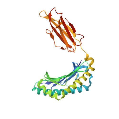
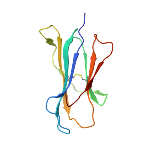
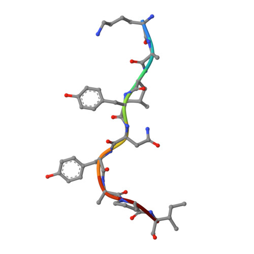
A structural basis for CD8+ T cell-dependent recognition of non-homologous peptide ligands: implications for molecular mimicry in autoreactivity

Sandalova, T.Michaelsson, J.Harris, R.A.Odeberg, J.Schneider, G.Karre, K.Achour, A.

(2005) J Biol Chem 280: 27069-27075

  • DOI: https://doi.org/10.1074/jbc.M500927200
  • Primary Citation of Related Structures:  
    1ZHB

  • PubMed Abstract: 

    Molecular mimicry of self-epitopes by viral antigens is one possible pathogenic mechanism underlying induction of autoimmunity. A self-epitope, mDBM, derived from mouse dopamine beta-mono-oxygenase (KALYDYAPI) sharing 44% sequence identity with the lymphocytic choriomeningitis virus-derived immunodominant epitope gp33 (KAVYNFATC/M), has previously been identified as a cross-reactive self-ligand, presentation of which results in autoimmunity. A rat peptide homologue, rDBM (KALYNYAPI, 56% identity to gp33), which displayed similar properties to mDBM, has also been identified. We herein report the crystal structure of H-2Db.rDBM and a comparison with the crystal structures of the cross-reactive H-2Db.gp33 and non-cross-reactive H-2Db.gp33 (V3L) escape variant (KALYNFATM, 88% identity to gp33). Despite the large sequence disparity, rDBM and gp33 peptides are presented in nearly identical manners by H-2Db, with a striking juxtaposition of the central sections of both peptides from residues p3 to p7. The structural similarity provides H-2Db in complex with either a virus-derived or a dopamine beta-mono-oxygenase-derived peptide with a shared antigenic identity that conserves the positioning of the heavy chain and peptide residues that interact with the T cell receptor (TCR). This stands in contrast to the structure of H-2Db.gp33 (V3L), in which a single conserved mutation, also present in rDBM, induces large movements of both the peptide backbone and the side chains that interact with the TCR. The TCR-interacting surfaces of the H-2Db.rDBM and H-2Db.gp33 major histocompatibility complexes are very similar with regard to shape, topology, and charge distribution, providing a structural basis for CD8 T cell activation by molecular mimicry and potential subsequent development of autoreactivity.


  • Organizational Affiliation

    Department of Medical Biochemistry and Biophysics, Microbiology and Tumor Biology Center, and Strategic Research Center IRIS for Studies of Integrated Recognition in the Immune System, Karolinska Institutet, SE-171 77 Stockholm, Sweden.


Macromolecules
Find similar proteins by:  (by identity cutoff)  |  3D Structure
Entity ID: 1
MoleculeChains Sequence LengthOrganismDetailsImage
H-2 class I histocompatibility antigen, D-B alpha chain
A, D, G, J
276Mus musculusMutation(s): 0 
Gene Names: H2-D
UniProt
Find proteins for P01899 (Mus musculus)
Explore P01899 
Go to UniProtKB:  P01899
Entity Groups  
Sequence Clusters30% Identity50% Identity70% Identity90% Identity95% Identity100% Identity
UniProt GroupP01899
Sequence Annotations
Expand
  • Reference Sequence
Find similar proteins by:  (by identity cutoff)  |  3D Structure
Entity ID: 2
MoleculeChains Sequence LengthOrganismDetailsImage
Beta-2-microglobulin
B, E, H, K
99Mus musculusMutation(s): 0 
Gene Names: B2M
UniProt & NIH Common Fund Data Resources
Find proteins for P01887 (Mus musculus)
Explore P01887 
Go to UniProtKB:  P01887
IMPC:  MGI:88127
Entity Groups  
Sequence Clusters30% Identity50% Identity70% Identity90% Identity95% Identity100% Identity
UniProt GroupP01887
Sequence Annotations
Expand
  • Reference Sequence

Find similar proteins by:  Sequence   |   3D Structure  

Entity ID: 3
MoleculeChains Sequence LengthOrganismDetailsImage
9-mer peptide from Dopamine beta-monooxygenase
C, F, I, L
9Rattus norvegicusMutation(s): 0 
EC: 1.14.17.1
UniProt
Find proteins for Q05754 (Rattus norvegicus)
Explore Q05754 
Go to UniProtKB:  Q05754
Entity Groups  
UniProt GroupQ05754
Sequence Annotations
Expand
  • Reference Sequence
Experimental Data & Validation

Experimental Data

  • Method: X-RAY DIFFRACTION
  • Resolution: 2.70 Å
  • R-Value Free: 0.262 
  • R-Value Work: 0.225 
  • R-Value Observed: 0.226 
  • Space Group: P 1 21 1
Unit Cell:
Length ( Å )Angle ( ˚ )
a = 92.063α = 90
b = 122.715β = 103
c = 99.403γ = 90
Software Package:
Software NamePurpose
REFMACrefinement
DENZOdata reduction
SCALEPACKdata scaling
AMoREphasing

Structure Validation

View Full Validation Report


Entry History 

Deposition Data

Revision History  (Full details and data files)

  • Version 1.0: 2005-06-14
    Type: Initial release
  • Version 1.1: 2008-04-30
    Changes: Version format compliance
  • Version 1.2: 2011-07-13
    Changes: Version format compliance
  • Version 1.3: 2017-11-29
    Changes: Data collection, Database references, Source and taxonomy
  • Version 1.4: 2023-10-25
    Changes: Data collection, Database references, Refinement description