Structural Determinants for Inhibitor Specificity and Selectivity in PDE2A Using the Wheat Germ in Vitro Translation System.
Iffland, A., Kohls, D., Low, S., Luan, J., Zhang, Y., Kothe, M., Cao, Q., Kamath, A.V., Ding, Y.H., Ellenberger, T.(2005) Biochemistry 44: 8312-8325
- PubMed: 15938621
- DOI: https://doi.org/10.1021/bi047313h
- Primary Citation of Related Structures:
1Z1L - PubMed Abstract:
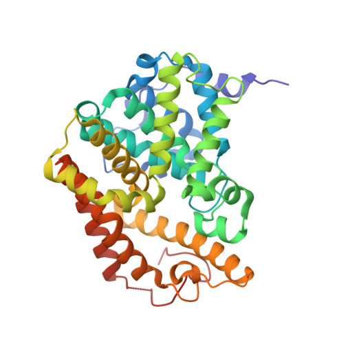
Phosphodiesterases (PDEs) modulate signaling by cyclic nucleotides in diverse processes such as cardiac contractility, platelet aggregation, lipolysis, glycogenolysis, and smooth muscle contraction. Cyclic guanosine monophosphate (cGMP) stimulated human phosphodiesterase 2 (PDE2) is expressed mainly in brain and heart tissues. PDE2A is involved in the regulation of blood pressure and fluid homeostasis by the atrial natriuretic peptide (ANP), making PDE2-type enzymes important targets for drug discovery. The design of more potent and selective inhibitors of PDE2A for the treatment of heart disease would be greatly aided by the identification of active site residues in PDE2A that determine substrate and inhibitor selectivity. The identification of active site residues through traditional mutational studies involves the time-consuming and tedious purification of a large number of mutant proteins from overexpressing cells. Here we report an alternative approach to rapidly produce active site mutants of human PDE2A and identify their enzymatic properties using a wheat germ in vitro translation (IVT, also known as cell-free translation) system. We also present the crystal structure of the catalytic domain of human PDE2A determined at 1.7 A resolution, which provided a framework for the rational design of active site mutants. Using a rapid IVT approach for expression of human PDE2A mutants, we identified the roles of active site residues Asp811, Gln812, Ile826, and Tyr827 in inhibitor and substrate selectivity for PDE2A.
Organizational Affiliation:
Department of Biology, Pfizer Research Technology Center, Cambridge, Massachusetts 02139, USA.