1YWG

The structure of glyceraldehyde-3-phosphate dehydrogenase from Plasmodium falciparum


Experimental Data Snapshot

  • Method: X-RAY DIFFRACTION
  • Resolution: 2.60 Å
  • R-Value Free: 0.256 
  • R-Value Work: 0.231 
  • R-Value Observed: 0.231 

wwPDB Validation   3D Report Full Report


Ligand Structure Quality Assessment 


This is version 1.4 of the entry. See complete history


Literature

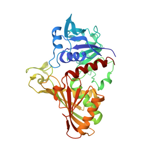
Structure of glyceraldehyde-3-phosphate dehydrogenase from Plasmodium falciparum.

Satchell, J.F.Malby, R.L.Luo, C.S.Adisa, A.Alpyurek, A.E.Klonis, N.Smith, B.J.Tilley, L.Colman, P.M.

(2005) Acta Crystallogr D Biol Crystallogr 61: 1213-1221

  • DOI: https://doi.org/10.1107/S0907444905018317
  • Primary Citation of Related Structures:  
    1YWG

  • PubMed Abstract: 

    The malaria parasite Plasmodium falciparum is responsible for about two million deaths annually, making it important to obtain information about enzymes from this organism that represent potential drug targets. The gene for P. falciparum glyceraldehyde-3-phosphate dehydrogenase (PfGAPDH) has been cloned and the protein expressed as a hexahistidine-tagged recombinant protein in Escherichia coli. The recombinant protein has been crystallized and its three-dimensional structure determined. One molecule of the cofactor NAD+ is bound to each of the four subunits in the tetrameric enzyme. The major structural feature distinguishing human GAPDH from PfGAPDH is the insertion of a dipeptide (-KG-) in the so-called S loop. This insert, together with other characteristic single-amino-acid substitutions, alters the chemical environment of the groove that encompasses the R dyad and that links adjacent cofactor-binding sites and may be responsible for the selective inhibition of the enzyme by ferriprotoporphyrin IX.


  • Organizational Affiliation

    The Walter and Eliza Hall Institute of Medical Research, Melbourne, Victoria, Australia.


Macromolecules
Find similar proteins by:  (by identity cutoff)  |  3D Structure
Entity ID: 1
MoleculeChains Sequence LengthOrganismDetailsImage
glyceraldehyde-3-phosphate dehydrogenaseA [auth O],
B [auth P],
C [auth Q],
D [auth R]
337Plasmodium falciparumMutation(s): 0 
Gene Names: 3D7
EC: 1.2.1.12
UniProt
Find proteins for Q8T6B1 (Plasmodium falciparum)
Explore Q8T6B1 
Go to UniProtKB:  Q8T6B1
Entity Groups  
Sequence Clusters30% Identity50% Identity70% Identity90% Identity95% Identity100% Identity
UniProt GroupQ8T6B1
Sequence Annotations
Expand
  • Reference Sequence
Experimental Data & Validation

Experimental Data

  • Method: X-RAY DIFFRACTION
  • Resolution: 2.60 Å
  • R-Value Free: 0.256 
  • R-Value Work: 0.231 
  • R-Value Observed: 0.231 
  • Space Group: P 61
Unit Cell:
Length ( Å )Angle ( ˚ )
a = 79.6α = 90
b = 79.6β = 90
c = 406.3γ = 120
Software Package:
Software NamePurpose
CNSrefinement
DENZOdata reduction
SCALEPACKdata scaling
MOLREPphasing

Structure Validation

View Full Validation Report


Ligand Structure Quality Assessment 


Entry History 

Deposition Data

Revision History  (Full details and data files)

  • Version 1.0: 2005-11-29
    Type: Initial release
  • Version 1.1: 2008-04-30
    Changes: Version format compliance
  • Version 1.2: 2011-07-13
    Changes: Version format compliance
  • Version 1.3: 2014-11-12
    Changes: Structure summary
  • Version 1.4: 2023-10-25
    Changes: Data collection, Database references, Derived calculations, Refinement description