1YU6

Crystal Structure of the Subtilisin Carlsberg:OMTKY3 Complex


Experimental Data Snapshot

  • Method: X-RAY DIFFRACTION
  • Resolution: 1.55 Å
  • R-Value Free: 0.205 
  • R-Value Work: 0.191 
  • R-Value Observed: 0.192 

wwPDB Validation   3D Report Full Report


This is version 1.3 of the entry. See complete history


Literature

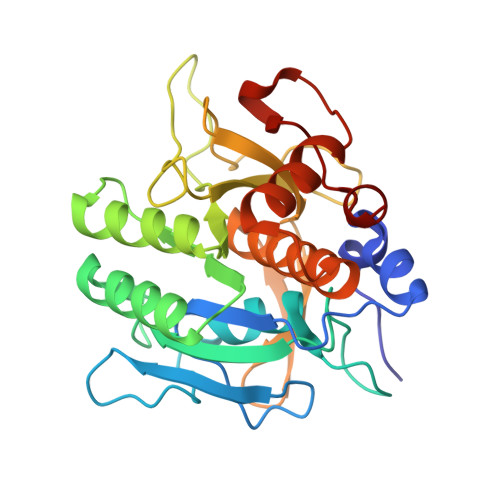
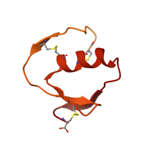
Structure of the subtilisin Carlsberg-OMTKY3 complex reveals two different ovomucoid conformations.

Maynes, J.T.Cherney, M.M.Qasim, M.A.Laskowski Jr, M.James, M.N.

(2005) Acta Crystallogr D Biol Crystallogr 61: 580-588

  • DOI: https://doi.org/10.1107/S0907444905004889
  • Primary Citation of Related Structures:  
    1YU6

  • PubMed Abstract: 

    One of the most studied protein proteinase inhibitors is the turkey ovomucoid third domain, OMTKY3. This inhibitor contains a reactive-site loop (Lys13I-Arg21I) that binds in a nearly identical manner to all studied serine proteinases, regardless of their clan or specificity. The crystal structure of OMTKY3 bound to subtilisin Carlsberg (CARL) has been determined. There are two complete copies of the complexes in the crystallographic asymmetric unit. Whereas the two enzyme molecules are virtually identical [0.16 A root-mean-square difference (r.m.s.d.) for 274 C(alpha) atoms], the two inhibitor molecules show dramatic differences between one another (r.m.s.d. = 2.4 A for 50 C(alpha) atoms). When compared with other proteinase-bound OMTKY3 molecules, these inhibitors show even larger differences. This work facilitates a re-evaluation of the importance of certain ovomucoid residues in proteinase binding and explains why additivity and sequence-based binding-prediction methods fail for the CARL-OMTKY3 complex.


  • Organizational Affiliation

    Canadian Institutes of Health Research, Group in Protein Structure and Function, Department of Biochemistry, Faculty of Medicine, University of Alberta, Edmonton, Alberta T6G 2H7, Canada. jason@biochem.ualberta.ca


Macromolecules
Find similar proteins by:  (by identity cutoff)  |  3D Structure
Entity ID: 1
MoleculeChains Sequence LengthOrganismDetailsImage
Subtilisin Carlsberg
A, B
275Bacillus licheniformisMutation(s): 0 
EC: 3.4.21.62
UniProt
Find proteins for P00780 (Bacillus licheniformis)
Explore P00780 
Go to UniProtKB:  P00780
Entity Groups  
Sequence Clusters30% Identity50% Identity70% Identity90% Identity95% Identity100% Identity
UniProt GroupP00780
Sequence Annotations
Expand
  • Reference Sequence
Find similar proteins by:  (by identity cutoff)  |  3D Structure
Entity ID: 2
MoleculeChains Sequence LengthOrganismDetailsImage
Ovomucoid
C, D
185Meleagris gallopavoMutation(s): 0 
UniProt
Find proteins for P68390 (Meleagris gallopavo)
Explore P68390 
Go to UniProtKB:  P68390
Entity Groups  
Sequence Clusters30% Identity50% Identity70% Identity90% Identity95% Identity100% Identity
UniProt GroupP68390
Sequence Annotations
Expand
  • Reference Sequence
Experimental Data & Validation

Experimental Data

  • Method: X-RAY DIFFRACTION
  • Resolution: 1.55 Å
  • R-Value Free: 0.205 
  • R-Value Work: 0.191 
  • R-Value Observed: 0.192 
  • Space Group: C 2 2 21
Unit Cell:
Length ( Å )Angle ( ˚ )
a = 110.256α = 90
b = 100.971β = 90
c = 115.785γ = 90
Software Package:
Software NamePurpose
REFMACrefinement
DENZOdata reduction
SCALEPACKdata scaling
AMoREphasing

Structure Validation

View Full Validation Report


Entry History 

Deposition Data

Revision History  (Full details and data files)

  • Version 1.0: 2005-05-03
    Type: Initial release
  • Version 1.1: 2008-04-30
    Changes: Version format compliance
  • Version 1.2: 2011-07-13
    Changes: Version format compliance
  • Version 1.3: 2023-08-23
    Changes: Data collection, Database references, Derived calculations, Refinement description