Structure and mode of action of the membrane-permeabilizing antimicrobial peptide pheromone plantaricin A
Kristiansen, P.E., Fimland, G., Mantzilas, D., Nissen-Meyer, J.(2005) J Biol Chem 280: 22945-22950
- PubMed: 15805109
- DOI: https://doi.org/10.1074/jbc.M501620200
- Primary Citation of Related Structures:
1YTR - PubMed Abstract:
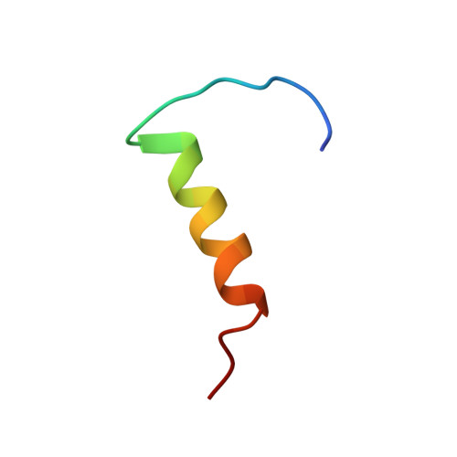
The three-dimensional structure in dodecyl phosphocholine micelles of the 26-mer membrane-permeabilizing bacteriocin-like pheromone plantaricin A (PlnA) has been determined by use of nuclear magnetic resonance spectroscopy. The peptide was unstructured in water but became partly structured upon exposure to micelles. An amphiphilic alpha-helix stretching from residue 12 to 21 (possibly also including residues 22 and 23) was then formed in the C-terminal part of the peptide, whereas the N-terminal part remained largely unstructured. PlnA exerted its membrane-permeabilizing antimicrobial activity through a nonchiral interaction with the target cell membrane because the d-enantiomeric form had the same activity as the natural l-form. This nonchiral interaction involved the amphiphilic alpha-helical region in the C-terminal half of PlnA because a 17-mer fragment that contains the amphiphilic alpha-helical part of the peptide had antimicrobial potency that was similar to that of the l- and d-enantiomeric forms of PlnA. Also the pheromone activity of PlnA depended on this nonchiral interaction because both the l- and d-enantiomeric forms of the 17-mer fragment inhibited the pheromone activity. The pheromone activity also involved, however, a chiral interaction between the N-terminal part of PlnA and its receptor because high concentrations of the l-form (but not the d-form) of a 5-mer fragment derived from the N-terminal part of PlnA had pheromone activity. The results thus reveal a novel mechanism whereby peptide pheromones such as PlnA may function. An initial nonchiral interaction with membrane lipids induces alpha-helical structuring in a segment of the peptide pheromone. The peptide becomes thereby sufficiently structured and properly positioned in the membrane interface, thus enabling it to engage in a chiral interaction with its receptor in or near the membrane water interface. This membrane-interacting mode of action explains why some peptide pheromones/hormones such as PlnA sometimes display antimicrobial activity in addition to their pheromone activity.
Organizational Affiliation:
Department of Molecular Biosciences, University of Oslo, Norway. eugen@kjemi.uio.no