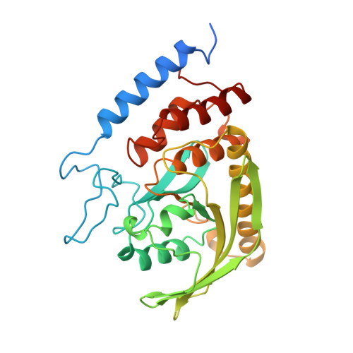
The X-ray crystal structures of Yersinia tyrosine phosphatase with bound tungstate and nitrate. Mechanistic implications.
Fauman, E.B., Yuvaniyama, C., Schubert, H.L., Stuckey, J.A., Saper, M.A.(1996) J Biol Chem 271: 18780-18788
- PubMed: 8702535
- DOI: https://doi.org/10.1074/jbc.271.31.18780
- Primary Citation of Related Structures:
1YTN, 1YTW - PubMed Abstract:
X-ray crystal structures of the Yersinia tyrosine phosphatase (PTPase) in complex with tungstate and nitrate have been solved to 2. 4-A resolution. Tetrahedral tungstate, WO42-, is a competitive inhibitor of the enzyme and is isosteric with the substrate and product of the catalyzed reaction. Planar nitrate, NO3-, is isosteric with the PO3 moiety of a phosphotransfer transition state. The crystal structures of the Yersinia PTPase with and without ligands, together with biochemical data, permit modeling of key steps along the reaction pathway. These energy-minimized models are consistent with a general acid-catalyzed, in-line displacement of the phosphate moiety to Cys403 on the enzyme, followed by attack by a nucleophilic water molecule to release orthophosphate. This nucleophilic water molecule is identified in the crystal structure of the nitrate complex. The active site structure of the PTPase is compared to alkaline phosphatase, which employs a similar phosphomonoester hydrolysis mechanism. Both enzymes must stabilize charges at the nucleophile, the PO3 moiety of the transition state, and the leaving group. Both an associative (bond formation preceding bond cleavage) and a dissociative (bond cleavage preceding bond formation) mechanism were modeled, but a dissociative-like mechanism is favored for steric and chemical reasons. Since nearly all of the 47 invariant or highly conserved residues of the PTPase domain are clustered at the active site, we suggest that the mechanism postulated for the Yersinia enzyme is applicable to all the PTPases.
Organizational Affiliation:
Biophysics Research Division and the Department of Biological Chemistry, The University of Michigan, Ann Arbor, Michigan 48109-1055, USA.