1YOO

ASPARTATE AMINOTRANSFERASE MUTANT ATB17 WITH ISOVALERIC ACID


Experimental Data Snapshot

  • Method: X-RAY DIFFRACTION
  • Resolution: 2.40 Å
  • R-Value Free: 0.235 
  • R-Value Work: 0.162 
  • R-Value Observed: 0.162 

wwPDB Validation   3D Report Full Report


This is version 1.4 of the entry. See complete history


Literature

Redesigning the substrate specificity of an enzyme by cumulative effects of the mutations of non-active site residues.

Oue, S.Okamoto, A.Yano, T.Kagamiyama, H.

(1999) J Biol Chem 274: 2344-2349

  • DOI: https://doi.org/10.1074/jbc.274.4.2344
  • Primary Citation of Related Structures:  
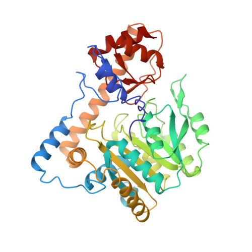
    1YOO

  • PubMed Abstract: 

    Directed evolution was used to change the substrate specificity of aspartate aminotransferase. A mutant enzyme with 17 amino acid substitutions was generated that shows a 2.1 x 10(6)-fold increase in the catalytic efficiency (kcat/Km) for a non-native substrate, valine. The absorption spectrum of the bound coenzyme, pyridoxal 5'-phosphate, is also changed significantly by the mutations. Interestingly, only one of the 17 residues appears to be able to contact the substrate, and none of them interact with the coenzyme. The three-dimensional structure of the mutant enzyme complexed with a valine analog, isovalerate (determined to 2.4-A resolution by x-ray crystallography), provides insights into how the mutations affect substrate binding. The active site is remodeled; the subunit interface is altered, and the enzyme domain that encloses the substrate is shifted by the mutations. The present results demonstrate clearly the importance of the cumulative effects of residues remote from the active site and represent a new line of approach to the redesign of enzyme activity.


  • Organizational Affiliation

    Department of Biochemistry, Osaka Medical College, Takatsuki, Osaka 569-8686, Japan.


Macromolecules
Find similar proteins by:  (by identity cutoff)  |  3D Structure
Entity ID: 1
MoleculeChains Sequence LengthOrganismDetailsImage
ASPARTATE AMINOTRANSFERASE396Escherichia coliMutation(s): 17 
EC: 2.6.1.1
UniProt
Find proteins for P00509 (Escherichia coli (strain K12))
Explore P00509 
Go to UniProtKB:  P00509
Entity Groups  
Sequence Clusters30% Identity50% Identity70% Identity90% Identity95% Identity100% Identity
UniProt GroupP00509
Sequence Annotations
Expand
  • Reference Sequence
Small Molecules
Ligands 2 Unique
IDChains Name / Formula / InChI Key2D Diagram3D Interactions
PLP
Query on PLP

Download Ideal Coordinates CCD File 
B [auth A]PYRIDOXAL-5'-PHOSPHATE
C8 H10 N O6 P
NGVDGCNFYWLIFO-UHFFFAOYSA-N
IVA
Query on IVA

Download Ideal Coordinates CCD File 
C [auth A]ISOVALERIC ACID
C5 H10 O2
GWYFCOCPABKNJV-UHFFFAOYSA-N
Experimental Data & Validation

Experimental Data

  • Method: X-RAY DIFFRACTION
  • Resolution: 2.40 Å
  • R-Value Free: 0.235 
  • R-Value Work: 0.162 
  • R-Value Observed: 0.162 
  • Space Group: C 2 2 21
Unit Cell:
Length ( Å )Angle ( ˚ )
a = 157α = 90
b = 84.84β = 90
c = 78.62γ = 90
Software Package:
Software NamePurpose
X-PLORmodel building
X-PLORrefinement
PROCESSdata reduction
PROCESSdata scaling
X-PLORphasing

Structure Validation

View Full Validation Report


Entry History 

Deposition Data

Revision History  (Full details and data files)

  • Version 1.0: 1999-02-02
    Type: Initial release
  • Version 1.1: 2008-03-24
    Changes: Version format compliance
  • Version 1.2: 2011-07-13
    Changes: Derived calculations, Version format compliance
  • Version 1.3: 2017-11-29
    Changes: Derived calculations, Other
  • Version 1.4: 2023-08-09
    Changes: Database references, Derived calculations, Refinement description