1YJC

SUBTILISIN BPN' 8397+1 (E.C. 3.4.21.14) (MUTANT WITH MET 50 REPLACED BY PHE, ASN 76 REPLACED BY ASP, GLY 169 REPLACED BY ALA, GLN 206 REPLACED BY CYS, ASN 218 REPLACED BY SER AND LYS 256 REPLACED BY TYR) (M50F, N76D, G169A, Q206C, N218S, AND K256Y) IN 50% DIMETHYLFORMAMIDE


Experimental Data Snapshot

  • Method: X-RAY DIFFRACTION
  • Resolution: 1.80 Å
  • R-Value Work: 0.187 
  • R-Value Observed: 0.187 

wwPDB Validation   3D Report Full Report


This is version 1.3 of the entry. See complete history


Literature

Breaking the low barrier hydrogen bond in a serine protease.

Kidd, R.D.Sears, P.Huang, D.H.Witte, K.Wong, C.H.Farber, G.K.

(1999) Protein Sci 8: 410-417

  • DOI: https://doi.org/10.1110/ps.8.2.410
  • Primary Citation of Related Structures:  
    1YJA, 1YJB, 1YJC

  • PubMed Abstract: 

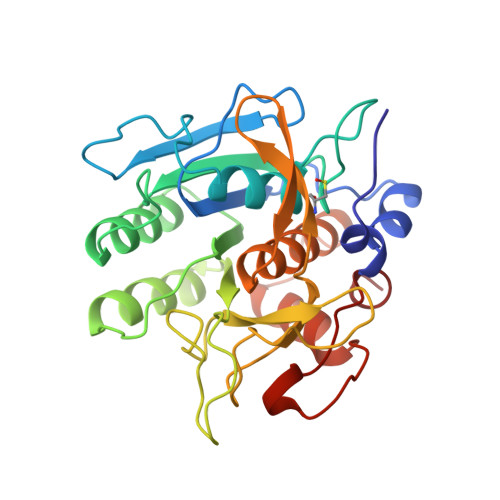
    The serine protease subtilisin BPN' is a useful catalyst for peptide synthesis when dissolved in high concentrations of a water-miscible organic co-solvent such as N,N-dimethylformamide (DMF). However, in 50% DMF, the k(cat) for amide hydrolysis is two orders of magnitude lower than in aqueous solution. Surprisingly, the k(cat) for ester hydrolysis is unchanged in 50% DMF. To explain this alteration in activity, the structure of subtilisin 8397+1 was determined in 20, 35, and 50% (v/v) DMF to 1.8 A resolution. In 50% DMF, the imidazole ring of His64, the central residue of the catalytic triad, has rotated approximately 180 degrees around the Cbeta-Cgamma bond. Two new water molecules in the active site stabilize the rotated conformation. This rotation places His64 in an unfavorable geometry to interact with the other members of the catalytic triad, Ser221 and Asp32. NMR experiments confirm that the characteristic resonance due to the low barrier hydrogen bond between the His64 and Asp32 is absent in 50% DMF. These experiments provide a clear structural basis for the change in activity of serine proteases in organic co-solvents.


  • Organizational Affiliation

    Department of Biochemistry and Molecular Biology, The Pennsylvania State University, University Park 16802-4500, USA.


Macromolecules
Find similar proteins by:  (by identity cutoff)  |  3D Structure
Entity ID: 1
MoleculeChains Sequence LengthOrganismDetailsImage
SUBTILISIN 8397+1275Bacillus amyloliquefaciensMutation(s): 6 
Gene Names: 8397+1
EC: 3.4.21.14
UniProt
Find proteins for P00782 (Bacillus amyloliquefaciens)
Explore P00782 
Go to UniProtKB:  P00782
Entity Groups  
Sequence Clusters30% Identity50% Identity70% Identity90% Identity95% Identity100% Identity
UniProt GroupP00782
Sequence Annotations
Expand
  • Reference Sequence
Small Molecules
Modified Residues  1 Unique
IDChains TypeFormula2D DiagramParent
CSO
Query on CSO
A
L-PEPTIDE LINKINGC3 H7 N O3 SCYS
Experimental Data & Validation

Experimental Data

  • Method: X-RAY DIFFRACTION
  • Resolution: 1.80 Å
  • R-Value Work: 0.187 
  • R-Value Observed: 0.187 
  • Space Group: P 1 21 1
Unit Cell:
Length ( Å )Angle ( ˚ )
a = 41.57α = 90
b = 79.7β = 114.2
c = 37.18γ = 90
Software Package:
Software NamePurpose
X-PLORmodel building
X-PLORrefinement
TEXSANdata reduction
X-PLORphasing

Structure Validation

View Full Validation Report


Entry History 

Deposition Data

Revision History  (Full details and data files)

  • Version 1.0: 1996-07-11
    Type: Initial release
  • Version 1.1: 2008-03-24
    Changes: Version format compliance
  • Version 1.2: 2011-07-13
    Changes: Version format compliance
  • Version 1.3: 2021-11-03
    Changes: Database references, Derived calculations, Other