1YDP

1.9A crystal structure of HLA-G


Experimental Data Snapshot

  • Method: X-RAY DIFFRACTION
  • Resolution: 1.90 Å
  • R-Value Free: 0.264 
  • R-Value Work: 0.235 

wwPDB Validation   3D Report Full Report


This is version 1.4 of the entry. See complete history


Literature

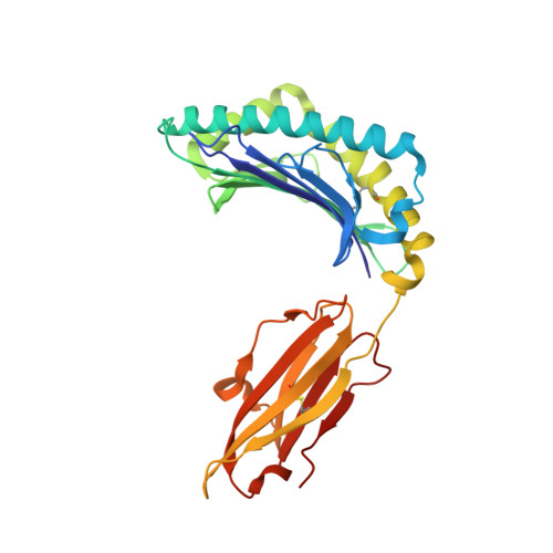
Crystal structure of HLA-G: A nonclassical MHC class I molecule expressed at the fetal-maternal interface

Clements, C.S.Kjer-Nielsen, L.Kostenko, L.Hoare, H.L.Dunstone, M.A.Moses, E.Freed, K.Brooks, A.G.Rossjohn, J.McCluskey, J.

(2005) Proc Natl Acad Sci U S A 102: 3360-3365

  • DOI: https://doi.org/10.1073/pnas.0409676102
  • Primary Citation of Related Structures:  
    1YDP

  • PubMed Abstract: 

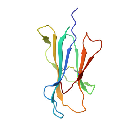
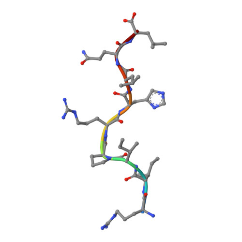
    HLA-G is a nonclassical major histocompatibility complex class I (MHC-I) molecule that is primarily expressed at the fetal-maternal interface, where it is thought to play a role in protecting the fetus from the maternal immune response. HLA-G binds a limited repertoire of peptides and interacts with the inhibitory leukocyte Ig-like receptors LIR-1 and LIR-2 and possibly with certain natural killer cell receptors. To gain further insights into HLA-G function, we determined the 1.9-A structure of a monomeric HLA-G complexed to a natural endogenous peptide ligand from histone H2A (RIIPRHLQL). An extensive network of contacts between the peptide and the antigen-binding cleft reveal a constrained mode of binding reminiscent of the nonclassical HLA-E molecule, thereby providing a structural basis for the limited peptide repertoire of HLA-G. The alpha3 domain of HLA-G, a candidate binding site for the LIR-1 and -2 inhibitory receptors, is structurally distinct from the alpha3 domains of classical MHC-I molecules, providing a rationale for the observed affinity differences for these ligands. The structural data suggest a head-to-tail mode of dimerization, mediated by an intermolecular disulfide bond, that is consistent with the observation of HLA-G dimers on the cell surface.


  • Organizational Affiliation

    Protein Crystallography Unit, Monash Centre for Synchrotron Science, Department of Biochemistry and Molecular Biology, School of Biomedical Sciences, Monash University, Clayton, Victoria 3800, Australia.


Macromolecules
Find similar proteins by:  (by identity cutoff)  |  3D Structure
Entity ID: 1
MoleculeChains Sequence LengthOrganismDetailsImage
MHC class I antigen275Homo sapiensMutation(s): 1 
UniProt & NIH Common Fund Data Resources
Find proteins for P17693 (Homo sapiens)
Explore P17693 
Go to UniProtKB:  P17693
PHAROS:  P17693
GTEx:  ENSG00000204632 
Entity Groups  
Sequence Clusters30% Identity50% Identity70% Identity90% Identity95% Identity100% Identity
UniProt GroupP17693
Sequence Annotations
Expand
  • Reference Sequence
Find similar proteins by:  (by identity cutoff)  |  3D Structure
Entity ID: 2
MoleculeChains Sequence LengthOrganismDetailsImage
Beta-2-microglobulin100Homo sapiensMutation(s): 0 
UniProt & NIH Common Fund Data Resources
Find proteins for P61769 (Homo sapiens)
Explore P61769 
Go to UniProtKB:  P61769
PHAROS:  P61769
GTEx:  ENSG00000166710 
Entity Groups  
Sequence Clusters30% Identity50% Identity70% Identity90% Identity95% Identity100% Identity
UniProt GroupP61769
Sequence Annotations
Expand
  • Reference Sequence

Find similar proteins by:  Sequence   |   3D Structure  

Entity ID: 3
MoleculeChains Sequence LengthOrganismDetailsImage
histone 2a peptideC [auth P]9synthetic constructMutation(s): 0 
UniProt & NIH Common Fund Data Resources
Find proteins for P16104 (Homo sapiens)
Explore P16104 
Go to UniProtKB:  P16104
PHAROS:  P16104
GTEx:  ENSG00000188486 
Entity Groups  
UniProt GroupP16104
Sequence Annotations
Expand
  • Reference Sequence
Small Molecules
Ligands 2 Unique
IDChains Name / Formula / InChI Key2D Diagram3D Interactions
CO
Query on CO

Download Ideal Coordinates CCD File 
D [auth B]COBALT (II) ION
Co
XLJKHNWPARRRJB-UHFFFAOYSA-N
CL
Query on CL

Download Ideal Coordinates CCD File 
E [auth B],
F [auth B]
CHLORIDE ION
Cl
VEXZGXHMUGYJMC-UHFFFAOYSA-M
Experimental Data & Validation

Experimental Data

  • Method: X-RAY DIFFRACTION
  • Resolution: 1.90 Å
  • R-Value Free: 0.264 
  • R-Value Work: 0.235 
  • Space Group: P 32 2 1
Unit Cell:
Length ( Å )Angle ( ˚ )
a = 77.15α = 90
b = 77.15β = 90
c = 151.72γ = 120
Software Package:
Software NamePurpose
CNSrefinement

Structure Validation

View Full Validation Report


Entry History 

Deposition Data

Revision History  (Full details and data files)

  • Version 1.0: 2005-03-08
    Type: Initial release
  • Version 1.1: 2008-04-30
    Changes: Version format compliance
  • Version 1.2: 2011-07-13
    Changes: Version format compliance
  • Version 1.3: 2018-07-25
    Changes: Data collection, Source and taxonomy, Structure summary
  • Version 1.4: 2021-11-10
    Changes: Database references, Derived calculations