1YCP

THE CRYSTAL STRUCTURE OF FIBRINOGEN-AA PEPTIDE 1-23 (F8Y) BOUND TO BOVINE THROMBIN EXPLAINS WHY THE MUTATION OF PHE-8 TO TYROSINE STRONGLY INHIBITS NORMAL CLEAVAGE AT ARGININE-16


Experimental Data Snapshot

  • Method: X-RAY DIFFRACTION
  • Resolution: 2.50 Å
  • R-Value Free: 0.245 
  • R-Value Work: 0.183 
  • R-Value Observed: 0.183 

wwPDB Validation   3D Report Full Report


This is version 1.3 of the entry. See complete history


Literature

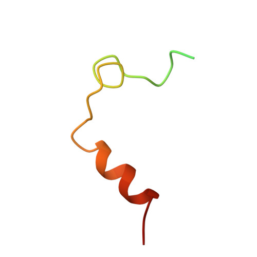
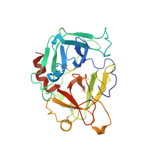
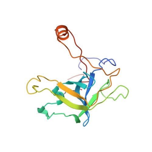
Crystal structure of fibrinogen-Aalpha peptide 1-23 (F8Y) bound to bovine thrombin explains why the mutation of Phe-8 to tyrosine strongly inhibits normal cleavage at Arg-16.

Malkowski, M.G.Martin, P.D.Lord, S.T.Edwards, B.F.

(1997) Biochem J 326: 815-822

  • DOI: https://doi.org/10.1042/bj3260815
  • Primary Citation of Related Structures:  
    1YCP

  • PubMed Abstract: 

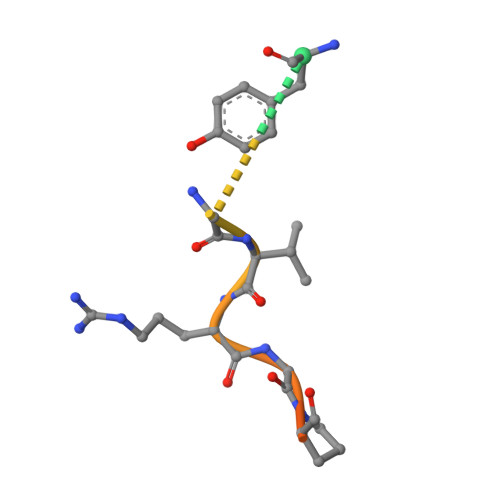
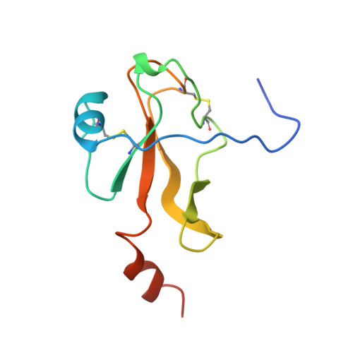
    A peptide containing residues 1-50 of the Aalpha-chain of fibrinogen, expressed as a fusion peptide with beta-galactosidase, is rapidly cleaved by thrombin at Arg-16, similarly to whole fibrinogen. When Phe-8, which is highly conserved, is replaced with tyrosine (F8Y), the cleavage is slowed drastically [Lord, Byrd, Hede, Wei and Colby (1990) J. Biol. Chem. 265, 838-843]. To examine the structural basis for this result, we have determined the crystal structure of bovine thrombin complexed with a synthetic peptide containing residues 1-23 of fibrinogen Aalpha and the F8Y mutation. The crystals are in space group P43212, with unit-cell dimensions of a = 88.3 A (1 A = 0.1 nm), c = 195.5 A and two complexes in the asymmetric unit. The final R factor is 0.183 for 2sigma data from 7.0 to 2.5 A resolution. There is continuous density for the five residues in the P3, P2, P1, P1' and P2' positions of the peptide (Gly-14f to Pro-18f) at the active site of thrombin, and isolated but well-defined density for Tyr-8f at position P9 in the hydrophobic pocket of thrombin. The tyrosine residue is shifted relative to phenylalanine in the native peptide because the phenol side chain is larger and makes a novel, intrapeptide hydrogen bond with Gly-14f. Adjacent peptide residues cannot form the hydrogen bonds that stabilize the secondary structure of the native peptide. Consequently, the 'reaction'geometry at the scissile bond, eight residues from the mutation, is perturbed and the peptide is mostly uncleaved in the crystal structure.


  • Organizational Affiliation

    Department of Biochemistry, Wayne State University, 540 E. Canfield Avenue, Detroit, MI 48201, USA.


Macromolecules
Find similar proteins by:  (by identity cutoff)  |  3D Structure
Entity ID: 1
MoleculeChains Sequence LengthOrganismDetailsImage
EPSILON THROMBINA [auth L],
D [auth J]
49Bos taurusMutation(s): 0 
EC: 3.4.21.5
UniProt
Find proteins for P00735 (Bos taurus)
Explore P00735 
Go to UniProtKB:  P00735
Entity Groups  
Sequence Clusters30% Identity50% Identity70% Identity90% Identity95% Identity100% Identity
UniProt GroupP00735
Sequence Annotations
Expand
  • Reference Sequence
Find similar proteins by:  (by identity cutoff)  |  3D Structure
Entity ID: 2
MoleculeChains Sequence LengthOrganismDetailsImage
ALPHA THROMBINB [auth H]259Bos taurusMutation(s): 0 
EC: 3.4.21.5
UniProt
Find proteins for P00735 (Bos taurus)
Explore P00735 
Go to UniProtKB:  P00735
Entity Groups  
Sequence Clusters30% Identity50% Identity70% Identity90% Identity95% Identity100% Identity
UniProt GroupP00735
Sequence Annotations
Expand
  • Reference Sequence

Find similar proteins by:  Sequence   |   3D Structure  

Entity ID: 3
MoleculeChains Sequence LengthOrganismDetailsImage
FIBRINOPEPTIDE A-ALPHAC [auth F],
G [auth N]
23Homo sapiensMutation(s): 0 
Gene Names: FGA
UniProt & NIH Common Fund Data Resources
Find proteins for P02671 (Homo sapiens)
Explore P02671 
Go to UniProtKB:  P02671
PHAROS:  P02671
GTEx:  ENSG00000171560 
Entity Groups  
Sequence Clusters30% Identity50% Identity70% Identity90% Identity95% Identity100% Identity
UniProt GroupP02671
Sequence Annotations
Expand
  • Reference Sequence
Find similar proteins by:  (by identity cutoff)  |  3D Structure
Entity ID: 4
MoleculeChains Sequence LengthOrganismDetailsImage
EPSILON THROMBINE [auth K]150Bos taurusMutation(s): 0 
UniProt
Find proteins for P00735 (Bos taurus)
Explore P00735 
Go to UniProtKB:  P00735
Entity Groups  
Sequence Clusters30% Identity50% Identity70% Identity90% Identity95% Identity100% Identity
UniProt GroupP00735
Sequence Annotations
Expand
  • Reference Sequence
Find similar proteins by:  (by identity cutoff)  |  3D Structure
Entity ID: 5
MoleculeChains Sequence LengthOrganismDetailsImage
EPSILON THROMBINF [auth M]109Bos taurusMutation(s): 1 
UniProt
Find proteins for P00735 (Bos taurus)
Explore P00735 
Go to UniProtKB:  P00735
Entity Groups  
Sequence Clusters30% Identity50% Identity70% Identity90% Identity95% Identity100% Identity
UniProt GroupP00735
Sequence Annotations
Expand
  • Reference Sequence
Experimental Data & Validation

Experimental Data

  • Method: X-RAY DIFFRACTION
  • Resolution: 2.50 Å
  • R-Value Free: 0.245 
  • R-Value Work: 0.183 
  • R-Value Observed: 0.183 
  • Space Group: P 43 21 2
Unit Cell:
Length ( Å )Angle ( ˚ )
a = 88.27α = 90
b = 88.27β = 90
c = 195.53γ = 90
Software Package:
Software NamePurpose
XENGENdata collection
XENGENdata reduction
X-PLORmodel building
X-PLORrefinement
XENGENdata scaling
X-PLORphasing

Structure Validation

View Full Validation Report


Entry History 

Deposition Data

Revision History  (Full details and data files)

  • Version 1.0: 1998-05-06
    Type: Initial release
  • Version 1.1: 2008-03-24
    Changes: Version format compliance
  • Version 1.2: 2011-07-13
    Changes: Atomic model, Database references, Derived calculations, Non-polymer description, Structure summary, Version format compliance
  • Version 1.3: 2021-11-03
    Changes: Database references, Other