1Y7V

X-ray structure of human acid-beta-glucosidase covalently bound to conduritol B epoxide


Experimental Data Snapshot

  • Method: X-RAY DIFFRACTION
  • Resolution: 2.40 Å
  • R-Value Free: 0.280 
  • R-Value Work: 0.242 
  • R-Value Observed: 0.244 

wwPDB Validation   3D Report Full Report


Ligand Structure Quality Assessment 


This is version 2.2 of the entry. See complete history


Literature

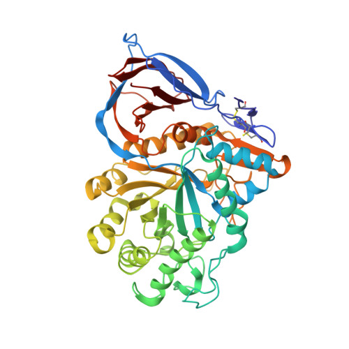
X-ray structure of human acid-beta-glucosidase covalently bound to conduritol-B-epoxide. Implications for Gaucher disease.

Premkumar, L.Sawkar, A.R.Boldin-Adamsky, S.Toker, L.Silman, I.Kelly, J.W.Futerman, A.H.Sussman, J.L.

(2005) J Biol Chem 280: 23815-23819

  • DOI: https://doi.org/10.1074/jbc.M502799200
  • Primary Citation of Related Structures:  
    1Y7V

  • PubMed Abstract: 

    Gaucher disease is an inherited metabolic disorder caused by mutations in the lysosomal enzyme acid-beta-glucosidase (GlcCerase). We recently determined the x-ray structure of GlcCerase to 2.0 A resolution (Dvir, H., Harel, M., McCarthy, A. A., Toker, L., Silman, I., Futerman, A. H., and Sussman, J. L. (2003) EMBO Rep.4, 704-709) and have now solved the structure of Glc-Cerase conjugated with an irreversible inhibitor, conduritol-B-epoxide (CBE). The crystal structure reveals that binding of CBE to the active site does not induce a global conformational change in GlcCerase and confirms that Glu340 is the catalytic nucleophile. However, only one of two alternative conformations of a pair of flexible loops (residues 345-349 and 394-399) located at the entrance to the active site in native GlcCerase is observed in the GlcCerase-CBE structure, a conformation in which the active site is accessible to CBE. Analysis of the dynamics of these two alternative conformations suggests that the two loops act as a lid at the entrance to the active site. This possibility is supported by a cluster of mutations in loop 394-399 that cause Gaucher disease by reducing catalytic activity. Moreover, in silico mutational analysis demonstrates that all these mutations stabilize the conformation that limits access to the active site, thus providing a mechanistic explanation of how mutations in this loop result in Gaucher disease.


  • Organizational Affiliation

    Department of Structural Biology, Weizmann Institute of Science, Rehovot 76100, Israel.


Macromolecules
Find similar proteins by:  (by identity cutoff)  |  3D Structure
Entity ID: 1
MoleculeChains Sequence LengthOrganismDetailsImage
Glucosylceramidase
A, B
497Homo sapiensMutation(s): 1 
Gene Names: GBAGC
EC: 3.2.1.45
UniProt & NIH Common Fund Data Resources
Find proteins for P04062 (Homo sapiens)
Explore P04062 
Go to UniProtKB:  P04062
PHAROS:  P04062
GTEx:  ENSG00000177628 
Entity Groups  
Sequence Clusters30% Identity50% Identity70% Identity90% Identity95% Identity100% Identity
UniProt GroupP04062
Sequence Annotations
Expand
  • Reference Sequence
Oligosaccharides

Help

Entity ID: 2
MoleculeChains Length2D Diagram Glycosylation3D Interactions
2-acetamido-2-deoxy-beta-D-glucopyranose-(1-4)-2-acetamido-2-deoxy-beta-D-glucopyranose
C, D
2N-Glycosylation
Glycosylation Resources
GlyTouCan:  G42666HT
GlyCosmos:  G42666HT
GlyGen:  G42666HT
Small Molecules
Ligands 2 Unique
IDChains Name / Formula / InChI Key2D Diagram3D Interactions
INS
Query on INS

Download Ideal Coordinates CCD File 
L [auth A],
S [auth B]
1,2,3,4,5,6-HEXAHYDROXY-CYCLOHEXANE
C6 H12 O6
CDAISMWEOUEBRE-GPIVLXJGSA-N
SO4
Query on SO4

Download Ideal Coordinates CCD File 
E [auth A]
F [auth A]
G [auth A]
H [auth A]
I [auth A]
E [auth A],
F [auth A],
G [auth A],
H [auth A],
I [auth A],
J [auth A],
K [auth A],
M [auth B],
N [auth B],
O [auth B],
P [auth B],
Q [auth B],
R [auth B]
SULFATE ION
O4 S
QAOWNCQODCNURD-UHFFFAOYSA-L
Experimental Data & Validation

Experimental Data

  • Method: X-RAY DIFFRACTION
  • Resolution: 2.40 Å
  • R-Value Free: 0.280 
  • R-Value Work: 0.242 
  • R-Value Observed: 0.244 
  • Space Group: C 2 2 21
Unit Cell:
Length ( Å )Angle ( ˚ )
a = 104.49α = 90
b = 285.61β = 90
c = 91.51γ = 90
Software Package:
Software NamePurpose
XDSdata scaling
XSCALEdata scaling
CNSrefinement
XDSdata reduction
CNSphasing

Structure Validation

View Full Validation Report


Ligand Structure Quality Assessment 


Entry History 

Revision History  (Full details and data files)

  • Version 1.0: 2005-04-12
    Type: Initial release
  • Version 1.1: 2008-04-30
    Changes: Version format compliance
  • Version 1.2: 2011-07-13
    Changes: Non-polymer description, Version format compliance
  • Version 2.0: 2020-07-29
    Type: Remediation
    Reason: Carbohydrate remediation
    Changes: Atomic model, Data collection, Derived calculations, Structure summary
  • Version 2.1: 2021-10-20
    Changes: Database references, Structure summary
  • Version 2.2: 2023-08-23
    Changes: Data collection, Refinement description