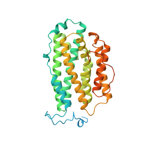
The three-dimensional structure of mammalian ribonucleotide reductase protein R2 reveals a more-accessible iron-radical site than Escherichia coli R2.
Kauppi, B., Nielsen, B.B., Ramaswamy, S., Larsen, I.K., Thelander, M., Thelander, L., Eklund, H.(1996) J Mol Biol 262: 706-720
- PubMed: 8876648
- DOI: https://doi.org/10.1006/jmbi.1996.0546
- Primary Citation of Related Structures:
1XSM - PubMed Abstract:
The three-dimensional structure of mouse ribonucleotide reductase R2 has been determined at 2.3 A resolution using molecular replacement and refined to an R-value of 19.1% (Rfree = 25%) with good stereo-chemistry. The overall tertiary structure architecture of mouse R2 is similar to that from Escherichia coli R2. However, several important structural differences are observed. Unlike the E. coli protein, the mouse dimer is completely devoid of beta-strands. The sequences differ significantly between the mouse and E. coli R2s, but there is high sequence identity among the eukaryotic R2 proteins, and the identities are localized over the whole sequence. Therefore, the three-dimensional structures of other mammalian ribonucleotide reductase R2 proteins are expected to be very similar to that of the mouse enzyme. In mouse R2 a narrow hydrophobic channel leads to the proposed binding site for molecular oxygen near to the iron-radical site in the interior of the protein. In E. coli R2 this channel is blocked by the phenyl ring of a tyrosine residue, which in mouse R2 is a serine. These structural variations may explain the observed differences in sensitivity to radical scavengers. The structure determination is based on diffraction data from crystals grown at pH 4.7. Unexpectedly, the protein is not iron-free, but contains one iron ion bound at one of the dinuclear iron sites. This ferric ion is bound with partial occupancy and is coordinated by three glutamic acids (one bidentate) and one histidine in a bipyramidal coordination that has a free apical coordination position. Soaking of crystals in a solution of ferrous salt at pH 4.7 increased the occupancy on the already occupied site, but without any detectable binding at the second site.
Organizational Affiliation:
Department of Molecular Biology, Swedish University of Agricultural Sciences, Biomedical Center, Uppsala, Sweden.