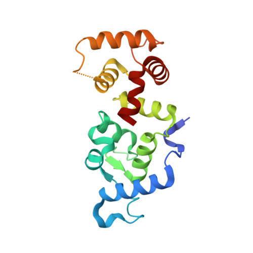
1XO5

Crystal structure of CIB1, an EF-hand, integrin and kinase-binding protein


Experimental Data Snapshot

  • Method: X-RAY DIFFRACTION
  • Resolution: 1.99 Å
  • R-Value Free: 0.257 
  • R-Value Work: 0.212 
  • R-Value Observed: 0.214 

wwPDB Validation   3D Report Full Report


This is version 1.4 of the entry. See complete history


Literature

Structural and Biochemical Characterization of CIB1 Delineates a New Family of EF-hand-containing Proteins

Gentry, H.R.Singer, A.U.Betts, L.Yang, C.Ferrara, J.D.Sondek, J.Parise, L.V.

(2005) J Biol Chem 280: 8407-8415

  • DOI: https://doi.org/10.1074/jbc.M411515200
  • Primary Citation of Related Structures:  
    1XO5

  • PubMed Abstract: 

    CIB1 (CIB) is an EF-hand-containing protein that binds multiple effector proteins, including the platelet alphaIIbbeta3 integrin and several serine/threonine kinases and potentially modulates their function. The crystal structure for Ca(2+)-bound CIB1 has been determined at 2.0 A resolution and reveals a compact alpha-helical protein containing four EF-hands, the last two of which bind calcium ions in the standard fashion seen in many other EF-hand proteins. CIB1 shares high structural similarity with calcineurin B and the neuronal calcium sensor (NCS) family of EF-hand-containing proteins. Most importantly, like calcineurin B and NCS proteins, which possess a large hydrophobic pocket necessary for ligand binding, CIB1 contains a hydrophobic pocket that has been implicated in ligand binding by previous mutational analysis. However, unlike several NCS proteins, Ca(2+)-bound CIB1 is largely monomeric whether bound to a relevant peptide ligand or ligand-free. Differences in structure, oligomeric state, and phylogeny define a new family of CIB1-related proteins that extends from arthropods to humans.


  • Organizational Affiliation

    Department of Pharmacology, The University of North Carolina, Chapel Hill 27599-7365, USA.


Macromolecules
Find similar proteins by:  (by identity cutoff)  |  3D Structure
Entity ID: 1
MoleculeChains Sequence LengthOrganismDetailsImage
Calcium and integrin-binding protein 1
A, B
183Homo sapiensMutation(s): 0 
Gene Names: CIB1PRKDCIPKIPCIB
UniProt & NIH Common Fund Data Resources
Find proteins for Q99828 (Homo sapiens)
Explore Q99828 
Go to UniProtKB:  Q99828
PHAROS:  Q99828
GTEx:  ENSG00000185043 
Entity Groups  
Sequence Clusters30% Identity50% Identity70% Identity90% Identity95% Identity100% Identity
UniProt GroupQ99828
Sequence Annotations
Expand
  • Reference Sequence
Experimental Data & Validation

Experimental Data

  • Method: X-RAY DIFFRACTION
  • Resolution: 1.99 Å
  • R-Value Free: 0.257 
  • R-Value Work: 0.212 
  • R-Value Observed: 0.214 
  • Space Group: P 1 21 1
Unit Cell:
Length ( Å )Angle ( ˚ )
a = 56.663α = 90
b = 51.009β = 103.12
c = 77.214γ = 90
Software Package:
Software NamePurpose
REFMACrefinement
MAR345data collection
SCALEPACKdata scaling
SOLVEphasing

Structure Validation

View Full Validation Report


Entry History 

Deposition Data

Revision History  (Full details and data files)

  • Version 1.0: 2004-12-07
    Type: Initial release
  • Version 1.1: 2008-04-30
    Changes: Version format compliance
  • Version 1.2: 2011-07-13
    Changes: Version format compliance
  • Version 1.3: 2017-10-11
    Changes: Refinement description
  • Version 1.4: 2024-02-14
    Changes: Data collection, Database references, Derived calculations