1XMM

Structure of human Dcps bound to m7GDP


Experimental Data Snapshot

  • Method: X-RAY DIFFRACTION
  • Resolution: 2.50 Å
  • R-Value Free: 0.267 
  • R-Value Work: 0.214 
  • R-Value Observed: 0.216 

wwPDB Validation   3D Report Full Report


Ligand Structure Quality Assessment 


This is version 2.0 of the entry. See complete history


Literature

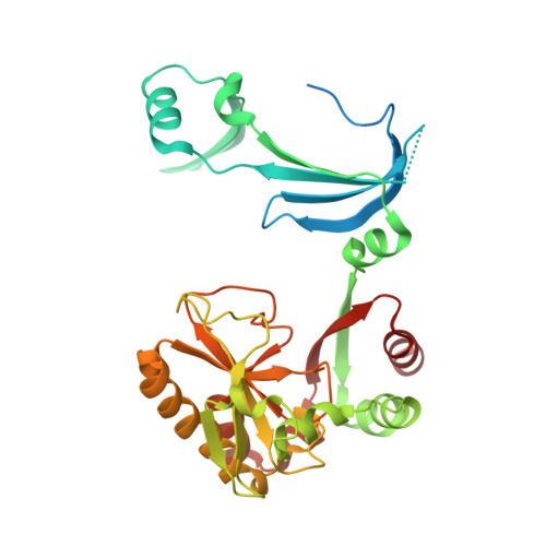
Crystal structures of human DcpS in ligand-free and m7GDP-bound forms suggest a dynamic mechanism for scavenger mRNA decapping.

Chen, N.Walsh, M.A.Liu, Y.Parker, R.Song, H.

(2005) J Mol Biol 347: 707-718

  • DOI: https://doi.org/10.1016/j.jmb.2005.01.062
  • Primary Citation of Related Structures:  
    1XML, 1XMM

  • PubMed Abstract: 

    Eukaryotic cells utilize DcpS, a scavenger decapping enzyme, to degrade the residual cap structure following 3'-5' mRNA decay, thereby preventing the premature decapping of the capped long mRNA and misincorporation of methylated nucleotides in nucleic acids. We report the structures of DcpS in ligand-free form and in a complex with m7GDP. apo-DcpS is a symmetric dimer, strikingly different from the asymmetric dimer observed in the structures of DcpS with bound cap analogues. In contrast, and similar to the m7GpppG-DcpS complex, DcpS with bound m7GDP is an asymmetric dimer in which the closed state appears to be the substrate-bound complex, whereas the open state mimics the product-bound complex. Comparisons of these structures revealed conformational changes of both the N-terminal swapped-dimeric domain and the cap-binding pocket upon cap binding. Moreover, Tyr273 in the cap-binding pocket displays remarkable conformational changes upon cap binding. Mutagenesis and biochemical analysis suggest that Tyr273 seems to play an important role in cap binding and product release. Examination of the crystallographic B-factors indicates that the N-terminal domain in apo-DcpS is inherently flexible, and in a dynamic state ready for substrate binding and product release.


  • Organizational Affiliation

    Laboratory of Macromolecular Structure, Institute of Molecular and Cell Biology, 61 Biopolis Drive, Proteos, Singapore 138673.


Macromolecules
Find similar proteins by:  (by identity cutoff)  |  3D Structure
Entity ID: 1
MoleculeChains Sequence LengthOrganismDetailsImage
heat shock-like protein 1A [auth B],
B [auth A],
C,
D
342Homo sapiensMutation(s): 0 
UniProt & NIH Common Fund Data Resources
Find proteins for Q96C86 (Homo sapiens)
Explore Q96C86 
Go to UniProtKB:  Q96C86
PHAROS:  Q96C86
GTEx:  ENSG00000110063 
Entity Groups  
Sequence Clusters30% Identity50% Identity70% Identity90% Identity95% Identity100% Identity
UniProt GroupQ96C86
Sequence Annotations
Expand
  • Reference Sequence
Small Molecules
Ligands 3 Unique
IDChains Name / Formula / InChI Key2D Diagram3D Interactions
M7G
Query on M7G

Download Ideal Coordinates CCD File 
H [auth A],
L [auth D]
7N-METHYL-8-HYDROGUANOSINE-5'-DIPHOSPHATE
C11 H18 N5 O11 P2
SBASPRRECYVBRF-KQYNXXCUSA-O
G7M
Query on G7M

Download Ideal Coordinates CCD File 
F [auth B],
J [auth C]
N7-METHYL-GUANOSINE-5'-MONOPHOSPHATE
C11 H17 N5 O8 P
AOKQNZVJJXPUQA-KQYNXXCUSA-O
PO4
Query on PO4

Download Ideal Coordinates CCD File 
E [auth B],
G [auth A],
I [auth C],
K [auth D]
PHOSPHATE ION
O4 P
NBIIXXVUZAFLBC-UHFFFAOYSA-K
Experimental Data & Validation

Experimental Data

  • Method: X-RAY DIFFRACTION
  • Resolution: 2.50 Å
  • R-Value Free: 0.267 
  • R-Value Work: 0.214 
  • R-Value Observed: 0.216 
  • Space Group: P 21 21 21
Unit Cell:
Length ( Å )Angle ( ˚ )
a = 100.928α = 90
b = 105.14β = 90
c = 138.647γ = 90
Software Package:
Software NamePurpose
REFMACrefinement
MOSFLMdata reduction
CCP4data scaling
MOLREPphasing

Structure Validation

View Full Validation Report


Ligand Structure Quality Assessment 


Entry History 

Deposition Data

Revision History  (Full details and data files)

  • Version 1.0: 2005-03-22
    Type: Initial release
  • Version 1.1: 2008-04-30
    Changes: Version format compliance
  • Version 1.2: 2011-07-13
    Changes: Advisory, Version format compliance
  • Version 2.0: 2024-03-06
    Changes: Data collection, Database references, Derived calculations, Non-polymer description, Structure summary