1XFO

Crystal Structure of an archaeal aminopeptidase


Experimental Data Snapshot

  • Method: X-RAY DIFFRACTION
  • Resolution: 1.96 Å
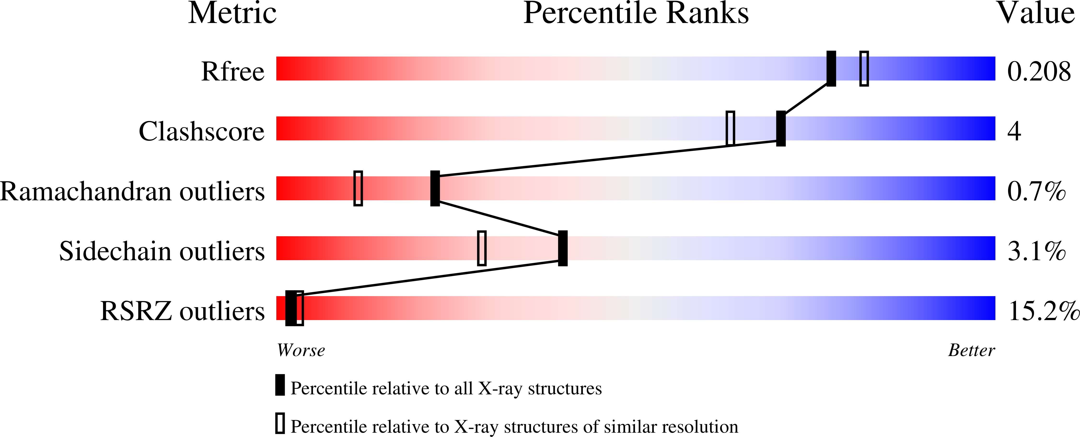
  • R-Value Free: 0.209 
  • R-Value Work: 0.183 
  • R-Value Observed: 0.183 

wwPDB Validation   3D Report Full Report


This is version 1.3 of the entry. See complete history


Literature

Crystal structure of a dodecameric tetrahedral shaped aminopeptidase

Russo, S.Baumann, U.

(2004) J Biol Chem 279: 51275-51281

  • DOI: https://doi.org/10.1074/jbc.M409455200
  • Primary Citation of Related Structures:  
    1XFO

  • PubMed Abstract: 

    Protein turnover is an essential process in living cells. The degradation of cytosolic polypeptides is mainly carried out by the proteasome, resulting in 7-9-amino acid long peptides. Further degradation is usually carried out by energy-independent proteases like the tricorn protease from Thermoplasma acidophilum. Recently, a novel tetrahedral-shaped dodecameric 480-kDa aminopeptidase complex (TET) has been described in Haloarcula marismortui that differs from the known ring- or barrel-shaped self-compartmentalizing proteases. This complex is capable of degrading most peptides down to amino acids. We present here the crystal structure of the tetrahedral aminopeptidase homolog FrvX from Pyrococcus horikoshii. The monomer has a typical clan MH fold, as found for example in Aeromonas proteolytica aminopeptidase, containing a dinuclear zinc active center. The quaternary structure is built by dimers with a length of 100 A that form the edges of the tetrahedron. All 12 active sites are located on the inside of the tetrahedron. Substrate access is granted by pores with a maximal diameter of 10 A, allowing only small peptides and unfolded proteins access to the active site.


  • Organizational Affiliation

    Departement für Chemie und Biochemie, University of Berne, Freiestrasse 3, CH-3012 Bern, Switzerland.


Macromolecules
Find similar proteins by:  (by identity cutoff)  |  3D Structure
Entity ID: 1
MoleculeChains Sequence LengthOrganismDetailsImage
Frv operon protein FrvX
A, B, C, D
357Pyrococcus horikoshiiMutation(s): 0 
UniProt
Find proteins for O59196 (Pyrococcus horikoshii (strain ATCC 700860 / DSM 12428 / JCM 9974 / NBRC 100139 / OT-3))
Explore O59196 
Go to UniProtKB:  O59196
Entity Groups  
Sequence Clusters30% Identity50% Identity70% Identity90% Identity95% Identity100% Identity
UniProt GroupO59196
Sequence Annotations
Expand
  • Reference Sequence
Experimental Data & Validation

Experimental Data

  • Method: X-RAY DIFFRACTION
  • Resolution: 1.96 Å
  • R-Value Free: 0.209 
  • R-Value Work: 0.183 
  • R-Value Observed: 0.183 
  • Space Group: P 63
Unit Cell:
Length ( Å )Angle ( ˚ )
a = 158.34α = 90
b = 158.34β = 90
c = 114.46γ = 120
Software Package:
Software NamePurpose
REFMACrefinement
MOSFLMdata reduction
XDSdata scaling
MOLREPphasing

Structure Validation

View Full Validation Report


Entry History 

Deposition Data

Revision History  (Full details and data files)

  • Version 1.0: 2004-10-05
    Type: Initial release
  • Version 1.1: 2008-04-30
    Changes: Version format compliance
  • Version 1.2: 2011-07-13
    Changes: Advisory, Derived calculations, Version format compliance
  • Version 1.3: 2023-10-25
    Changes: Data collection, Database references, Derived calculations, Refinement description