1XCB

X-ray Structure of a Rex-Family Repressor/NADH Complex from Thermus Aquaticus


Experimental Data Snapshot

  • Method: X-RAY DIFFRACTION
  • Resolution: 2.90 Å
  • R-Value Free: 0.276 
  • R-Value Work: 0.228 
  • R-Value Observed: 0.232 

wwPDB Validation   3D Report Full Report


Ligand Structure Quality Assessment 


This is version 1.3 of the entry. See complete history


Literature

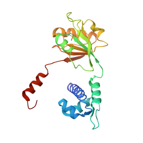
X-Ray Structure of a Rex-Family Repressor/NADH Complex: Insights into the Mechanism of Redox Sensing

Sickmier, E.A.Brekasis, D.Paranawithana, S.Bonanno, J.B.Paget, M.S.Burley, S.K.Kielkopf, C.L.

(2005) Structure 13: 43-54

  • DOI: https://doi.org/10.1016/j.str.2004.10.012
  • Primary Citation of Related Structures:  
    1XCB

  • PubMed Abstract: 

    The redox-sensing repressor Rex regulates transcription of respiratory genes in response to the intra cellular NADH/NAD(+) redox poise. As a step toward elucidating the molecular mechanism of NADH/NAD(+) sensing, the X-ray structure of Thermus aquaticus Rex (T-Rex) bound to effector NADH has been determined at 2.9 A resolution. The fold of the C-terminal domain of T-Rex is characteristic of NAD(H)-dependent enzymes, whereas the N-terminal domain is similar to a winged helix DNA binding motif. T-Rex dimerization is primarily mediated by "domain-swapped" alpha helices. Each NADH molecule binds to the C-terminal domain near the dimer interface. In contrast to NAD(H)-dependent enzymes, the nicotinamide is deeply buried within a hydrophobic pocket that appears to preclude substrate entry. We show that T-Rex binds to the Rex operator, and NADH but not NAD(+) inhibits T-Rex/DNA binding activity. A mechanism for redox sensing by Rex family members is proposed by analogy with domain closure of NAD(H)-dependent enzymes.


  • Organizational Affiliation

    Department of Biochemistry and Molecular Biology, Johns Hopkins Bloomberg School of Public Health, Baltimore, Maryland 21205, USA.


Macromolecules
Find similar proteins by:  (by identity cutoff)  |  3D Structure
Entity ID: 1
MoleculeChains Sequence LengthOrganismDetailsImage
Redox-sensing transcriptional repressor rex
A, B, C, D, E
A, B, C, D, E, F, G
211Thermus aquaticusMutation(s): 4 
Gene Names: rex
UniProt
Find proteins for Q9X2V5 (Thermus aquaticus)
Explore Q9X2V5 
Go to UniProtKB:  Q9X2V5
Entity Groups  
Sequence Clusters30% Identity50% Identity70% Identity90% Identity95% Identity100% Identity
UniProt GroupQ9X2V5
Sequence Annotations
Expand
  • Reference Sequence
Small Molecules
Ligands 2 Unique
IDChains Name / Formula / InChI Key2D Diagram3D Interactions
NAD
Query on NAD

Download Ideal Coordinates CCD File 
I [auth A]
K [auth B]
M [auth C]
P [auth D]
Q [auth E]
I [auth A],
K [auth B],
M [auth C],
P [auth D],
Q [auth E],
R [auth F],
S [auth G]
NICOTINAMIDE-ADENINE-DINUCLEOTIDE
C21 H27 N7 O14 P2
BAWFJGJZGIEFAR-NNYOXOHSSA-N
CA
Query on CA

Download Ideal Coordinates CCD File 
H [auth A],
J [auth B],
L [auth C],
N [auth D],
O [auth D]
CALCIUM ION
Ca
BHPQYMZQTOCNFJ-UHFFFAOYSA-N
Modified Residues  1 Unique
IDChains TypeFormula2D DiagramParent
MSE
Query on MSE
A, B, C, D, E
A, B, C, D, E, F, G
L-PEPTIDE LINKINGC5 H11 N O2 SeMET
Experimental Data & Validation

Experimental Data

  • Method: X-RAY DIFFRACTION
  • Resolution: 2.90 Å
  • R-Value Free: 0.276 
  • R-Value Work: 0.228 
  • R-Value Observed: 0.232 
  • Space Group: C 1 2 1
Unit Cell:
Length ( Å )Angle ( ˚ )
a = 189.24α = 90
b = 88.97β = 106.09
c = 112.91γ = 90
Software Package:
Software NamePurpose
DENZOdata reduction
SCALEPACKdata scaling
SOLVEphasing
CNSrefinement

Structure Validation

View Full Validation Report


Ligand Structure Quality Assessment 


Entry History 

Revision History  (Full details and data files)

  • Version 1.0: 2004-09-28
    Type: Initial release
  • Version 1.1: 2008-04-30
    Changes: Version format compliance
  • Version 1.2: 2011-07-13
    Changes: Version format compliance
  • Version 1.3: 2021-02-03
    Changes: Database references, Derived calculations, Structure summary