1X9Z

Crystal structure of the MutL C-terminal domain


Experimental Data Snapshot

  • Method: X-RAY DIFFRACTION
  • Resolution: 2.10 Å
  • R-Value Free: 0.278 
  • R-Value Work: 0.236 
  • R-Value Observed: 0.236 

wwPDB Validation   3D Report Full Report


This is version 1.2 of the entry. See complete history


Literature

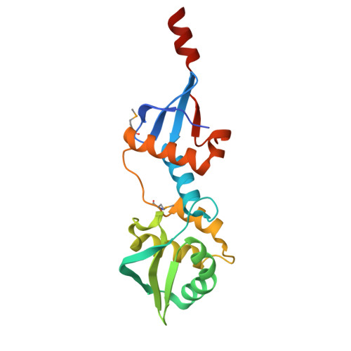
Structure of the MutL C-terminal domain: a model of intact MutL and its roles in mismatch repair

Guarne, A.Ramon-Maiques, S.Wolff, E.M.Ghirlando, R.Hu, X.Miller, J.H.Yang, W.

(2004) EMBO J 23: 4134-4145

  • DOI: https://doi.org/10.1038/sj.emboj.7600412
  • Primary Citation of Related Structures:  
    1X9Z

  • PubMed Abstract: 

    MutL assists the mismatch recognition protein MutS to initiate and coordinate mismatch repair in species ranging from bacteria to humans. The MutL N-terminal ATPase domain is highly conserved, but the C-terminal region shares little sequence similarity among MutL homologs. We report here the crystal structure of the Escherichia coli MutL C-terminal dimerization domain and the likelihood of its conservation among MutL homologs. A 100-residue proline-rich linker between the ATPase and dimerization domains, which generates a large central cavity in MutL dimers, tolerates sequence substitutions and deletions of one-third of its length with no functional consequences in vivo or in vitro. Along the surface of the central cavity, residues essential for DNA binding are located in both the N- and C-terminal domains. Each domain of MutL interacts with UvrD helicase and is required for activating the helicase activity. The DNA-binding capacity of MutL is correlated with the level of UvrD activation. A model of how MutL utilizes its ATPase and DNA-binding activities to mediate mismatch-dependent activation of MutH endonuclease and UvrD helicase is proposed.


  • Organizational Affiliation

    Laboratory of Molecular Biology, National Institute of Diabetes and Digestive and Kidney Diseases, National Institutes of Health, 9000 Rockville Pike, Bethesda, MD 20892, USA.


Macromolecules
Find similar proteins by:  (by identity cutoff)  |  3D Structure
Entity ID: 1
MoleculeChains Sequence LengthOrganismDetailsImage
DNA mismatch repair protein mutL
A, B
188Escherichia coliMutation(s): 2 
Gene Names: mutL
UniProt
Find proteins for P23367 (Escherichia coli (strain K12))
Explore P23367 
Go to UniProtKB:  P23367
Entity Groups  
Sequence Clusters30% Identity50% Identity70% Identity90% Identity95% Identity100% Identity
UniProt GroupP23367
Sequence Annotations
Expand
  • Reference Sequence
Small Molecules
Ligands 4 Unique
IDChains Name / Formula / InChI Key2D Diagram3D Interactions
GOL
Query on GOL

Download Ideal Coordinates CCD File 
F [auth A],
H [auth B]
GLYCEROL
C3 H8 O3
PEDCQBHIVMGVHV-UHFFFAOYSA-N
IPA
Query on IPA

Download Ideal Coordinates CCD File 
G [auth A],
I [auth B]
ISOPROPYL ALCOHOL
C3 H8 O
KFZMGEQAYNKOFK-UHFFFAOYSA-N
CL
Query on CL

Download Ideal Coordinates CCD File 
C [auth A],
D [auth A]
CHLORIDE ION
Cl
VEXZGXHMUGYJMC-UHFFFAOYSA-M
NA
Query on NA

Download Ideal Coordinates CCD File 
E [auth A]SODIUM ION
Na
FKNQFGJONOIPTF-UHFFFAOYSA-N
Modified Residues  1 Unique
IDChains TypeFormula2D DiagramParent
MSE
Query on MSE
A, B
L-PEPTIDE LINKINGC5 H11 N O2 SeMET
Experimental Data & Validation

Experimental Data

  • Method: X-RAY DIFFRACTION
  • Resolution: 2.10 Å
  • R-Value Free: 0.278 
  • R-Value Work: 0.236 
  • R-Value Observed: 0.236 
  • Space Group: P 43 2 2
Unit Cell:
Length ( Å )Angle ( ˚ )
a = 95.992α = 90
b = 95.992β = 90
c = 140.213γ = 90
Software Package:
Software NamePurpose
CNSrefinement
HKL-2000data reduction
SCALEPACKdata scaling
SOLVEphasing

Structure Validation

View Full Validation Report


Entry History 

Deposition Data

Revision History  (Full details and data files)

  • Version 1.0: 2004-10-26
    Type: Initial release
  • Version 1.1: 2008-04-30
    Changes: Version format compliance
  • Version 1.2: 2011-07-13
    Changes: Derived calculations, Version format compliance