1X8R

EPSPS liganded with the (S)-phosphonate analog of the tetrahedral reaction intermediate


Experimental Data Snapshot

  • Method: X-RAY DIFFRACTION
  • Resolution: 1.50 Å
  • R-Value Free: 0.195 
  • R-Value Work: 0.168 
  • R-Value Observed: 0.168 

wwPDB Validation   3D Report Full Report


Ligand Structure Quality Assessment 


This is version 1.4 of the entry. See complete history


Literature

Interaction of phosphonate analogues of the tetrahedral reaction intermediate with 5-enolpyruvylshikimate-3-phosphate synthase in atomic detail.

Priestman, M.A.Healy, M.L.Becker, A.Alberg, D.G.Bartlett, P.A.Lushington, G.H.Schonbrunn, E.

(2005) Biochemistry 44: 3241-3248

  • DOI: https://doi.org/10.1021/bi048198d
  • Primary Citation of Related Structures:  
    1X8R, 1X8T

  • PubMed Abstract: 

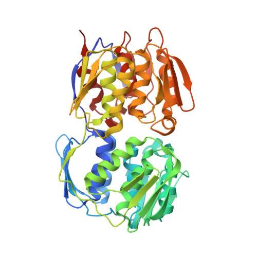
    The enzyme 5-enolpyruvylshikimate-3-phosphate synthase (EPSPS) catalyzes the penultimate step of the shikimate pathway and is the target of the broad-spectrum herbicide glyphosate. Since the functionality of the shikimate pathway is vital not only for plants but also for microorganisms, EPSPS is considered a prospective target for the development of novel antibiotics. We have kinetically analyzed and determined the crystal structures of Escherichia coli EPSPS inhibited by (R)- and (S)-configured phosphonate analogues of the tetrahedral reaction intermediate. Both diastereomers are competitive inhibitors with respect to the substrates of the EPSPS reaction, shikimate-3-phosphate (S3P) and phosphoenolpyruvate (PEP). Remarkably, the (S)-phosphonate (K(iS3P) = 750 nM), whose configuration corresponds to that of the genuine tetrahedral intermediate, is a much weaker inhibitor than the (R)-phosphonate analogue (K(iS3P) = 16 nM). The crystal structures of EPSPS liganded with the (S)- and (R)-phosphonates, at 1.5 and 1.9 A resolution, respectively, revealed that binding of the (R)-phosphonate induces conformational changes of the strictly conserved residues Arg124 and Glu341 within the active site. This appears to give rise to substantial structural alterations in the amino-terminal globular domain of the enzyme. By contrast, binding of the (S)-phosphonate renders the enzyme structure unchanged. Thus, EPSPS may facilitate the tight binding of structurally diverse ligands through conformational flexibility. Molecular docking calculations did not explain why the (R)-phosphonate is the better inhibitor. Therefore, we propose that the structural events during the open-closed transition of EPSPS are altered as a result of inhibitor action.


  • Organizational Affiliation

    Department of Medicinal Chemistry, University of Kansas, Lawrence, Kansas 66045, USA.


Macromolecules
Find similar proteins by:  (by identity cutoff)  |  3D Structure
Entity ID: 1
MoleculeChains Sequence LengthOrganismDetailsImage
3-phosphoshikimate 1-carboxyvinyltransferase427Escherichia coliMutation(s): 0 
Gene Names: aroA
EC: 2.5.1.19
UniProt
Find proteins for P0A6D3 (Escherichia coli (strain K12))
Explore P0A6D3 
Go to UniProtKB:  P0A6D3
Entity Groups  
Sequence Clusters30% Identity50% Identity70% Identity90% Identity95% Identity100% Identity
UniProt GroupP0A6D3
Sequence Annotations
Expand
  • Reference Sequence
Small Molecules
Ligands 2 Unique
IDChains Name / Formula / InChI Key2D Diagram3D Interactions
SC1
Query on SC1

Download Ideal Coordinates CCD File 
B [auth A][3R-[3A,4A,5B(S*)]]-5-(1-CARBOXY-1-PHOSPHONOETHOXY)-4-HYDROXY-3-(PHOSPHONOOXY)-1-CYCLOHEXENE-1-CARBOXYLIC ACID
C10 H16 O13 P2
HUOJJMMXOWLGJU-JQCUSGDOSA-N
FMT
Query on FMT

Download Ideal Coordinates CCD File 
C [auth A]
D [auth A]
E [auth A]
F [auth A]
G [auth A]
C [auth A],
D [auth A],
E [auth A],
F [auth A],
G [auth A],
H [auth A],
I [auth A],
J [auth A],
K [auth A],
L [auth A],
M [auth A],
N [auth A]
FORMIC ACID
C H2 O2
BDAGIHXWWSANSR-UHFFFAOYSA-N
Binding Affinity Annotations 
IDSourceBinding Affinity
SC1 PDBBind:  1X8R Ki: 750 (nM) from 1 assay(s)
Binding MOAD:  1X8R Ki: 2900 (nM) from 1 assay(s)
BindingDB:  1X8R Ki: 15 (nM) from 1 assay(s)
Kd: 15 (nM) from 1 assay(s)
Experimental Data & Validation

Experimental Data

  • Method: X-RAY DIFFRACTION
  • Resolution: 1.50 Å
  • R-Value Free: 0.195 
  • R-Value Work: 0.168 
  • R-Value Observed: 0.168 
  • Space Group: P 21 21 21
Unit Cell:
Length ( Å )Angle ( ˚ )
a = 57.73α = 90
b = 85.17β = 90
c = 87.49γ = 90
Software Package:
Software NamePurpose
XDSdata reduction
CNSrefinement
XDSdata scaling
CNSphasing

Structure Validation

View Full Validation Report


Ligand Structure Quality Assessment 


Entry History 

Deposition Data

Revision History  (Full details and data files)

  • Version 1.0: 2005-04-19
    Type: Initial release
  • Version 1.1: 2008-04-30
    Changes: Version format compliance
  • Version 1.2: 2011-07-13
    Changes: Version format compliance
  • Version 1.3: 2017-10-11
    Changes: Refinement description
  • Version 1.4: 2023-08-23
    Changes: Data collection, Database references, Derived calculations, Refinement description