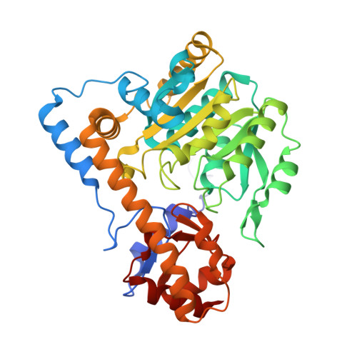
Binding of C5-dicarboxylic substrate to aspartate aminotransferase: implications for the conformational change at the transaldimination step.
Islam, M.M., Goto, M., Miyahara, I., Ikushiro, H., Hirotsu, K., Hayashi, H.(2005) Biochemistry 44: 8218-8229
- PubMed: 15938611
- DOI: https://doi.org/10.1021/bi050071g
- Primary Citation of Related Structures:
1X28, 1X29, 1X2A - PubMed Abstract:
The mechanism for the reaction of aspartate aminotransferase with the C4 substrate, l-aspartate, has been well established. The binding of the C4 substrate induces conformational change in the enzyme from the open to the closed form, and the entire reaction proceeds in the closed form of the enzyme. On the contrary, little is known about the reaction with the C5 substrate, l-glutamate. In this study, we analyzed the pH-dependent binding of 2-methyl-l-glutamate to the enzyme and showed that the interaction between the amino group of 2-methyl-l-glutamate and the pyridoxal 5'-phosphate aldimine is weak compared to that between 2-methyl-l-aspartate and the aldimine. The structures of the Michaelis complexes of the enzyme with l-aspartate and l-glutamate were modeled on the basis of the maleate and glutarate complex structures of the enzyme. The result showed that l-glutamate binds to the open form of the enzyme in an extended conformation, and its alpha-amino group points in the opposite direction of the aldimine, while that of l-aspartate is close to the aldimine. These models explain the observations for 2-methyl-l-glutamate and 2-methyl-l-aspartate. The crystal structures of the complexes of aspartate aminotransferase with phosphopyridoxyl derivatives of l-glutamate, d-glutamate, and 2-methyl-l-glutamate were solved as the models for the external aldimine and ketimine complexes of l-glutamate. All the structures were in the closed form, and the two carboxylate groups and the arginine residues binding them are superimposable on the external aldimine complex with 2-methyl-l-aspartate. Taking these facts altogether, it was strongly suggested that the binding of l-glutamate to aspartate aminotransferase to form the Michaelis complex does not induce a conformational change in the enzyme, and that the conformational change to the closed form occurs during the transaldimination step. The hydrophobic residues of the entrance of the active site, including Tyr70, are considered to be important for promoting the transaldimination process and hence the recognition of the C5 substrate.
Organizational Affiliation:
Department of Biochemistry, Osaka Medical College, 2-7 Daigakumachi, Takatsuki 569-8686, Japan.