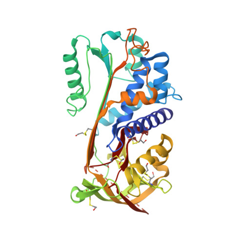
The high resolution crystal structure of the human tumor suppressor maspin reveals a novel conformational switch in the G-helix.
Law, R.H., Irving, J.A., Buckle, A.M., Ruzyla, K., Buzza, M., Bashtannyk-Puhalovich, T.A., Beddoe, T.C., Nguyen, K., Worrall, D.M., Bottomley, S.P., Bird, P.I., Rossjohn, J., Whisstock, J.C.(2005) J Biol Chem 280: 22356-22364
- PubMed: 15760906
- DOI: https://doi.org/10.1074/jbc.M412043200
- Primary Citation of Related Structures:
1WZ9, 1XU8 - PubMed Abstract:
Maspin is a serpin that acts as a tumor suppressor in a range of human cancers, including tumors of the breast and lung. Maspin is crucial for development, because homozygous loss of the gene is lethal; however, the precise physiological role of the molecule is unclear. To gain insight into the function of human maspin, we have determined its crystal structure in two similar, but non-isomorphous crystal forms, to 2.1- and 2.8-A resolution, respectively. The structure reveals that maspin adopts the native serpin fold in which the reactive center loop is expelled fully from the A beta-sheet, makes minimal contacts with the core of the molecule, and exhibits a high degree of flexibility. A buried salt bridge unique to maspin orthologues causes an unusual bulge in the region around the D and E alpha-helices, an area of the molecule demonstrated in other serpins to be important for cofactor recognition. Strikingly, the structural data reveal that maspin is able to undergo conformational change in and around the G alpha-helix, switching between an open and a closed form. This change dictates the electrostatic character of a putative cofactor binding surface and highlights this region as a likely determinant of maspin function. The high resolution crystal structure of maspin provides a detailed molecular framework to elucidate the mechanism of function of this important tumor suppressor.
Organizational Affiliation:
The Protein Crystallography Unit, Monash Centre for Synchrotron Science and The Department of Biochemistry and Molecular Biology, School of Biomedical Sciences, Faculty of Medicine, Monash University, Clayton, Victoria 3800, Australia.