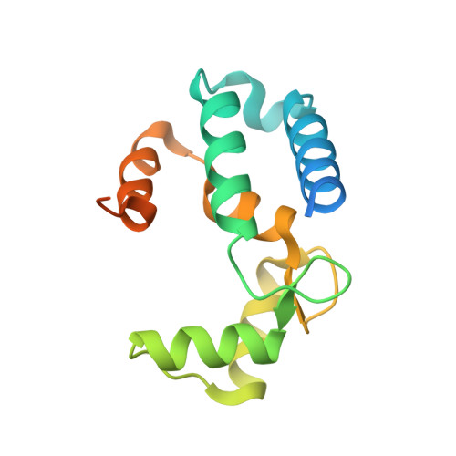
1WY9

Crystal structure of microglia-specific protein, Iba1


Experimental Data Snapshot

  • Method: X-RAY DIFFRACTION
  • Resolution: 2.10 Å
  • R-Value Free: 0.251 
  • R-Value Work: 0.217 
  • R-Value Observed: 0.217 

wwPDB Validation   3D Report Full Report


This is version 1.3 of the entry. See complete history


Literature

X-ray Structures of the Microglia/Macrophage-specific Protein Iba1 from Human and Mouse Demonstrate Novel Molecular Conformation Change Induced by Calcium binding

Yamada, M.Ohsawa, K.Imai, Y.Kohsaka, S.Kamitori, S.

(2006) J Mol Biol 364: 449-457

  • DOI: https://doi.org/10.1016/j.jmb.2006.09.027
  • Primary Citation of Related Structures:  
    1WY9, 2D58

  • PubMed Abstract: 

    The ionized calcium-binding adaptor molecule 1 (Iba1) with 147 amino acid residues has been identified as a calcium-binding protein, expressed specifically in microglia/macrophages, and is expected to be a key factor in membrane ruffling, which is a typical feature of activated microglia. We have determined the crystal structure of human Iba1 in a Ca(2+)-free form and mouse Iba1 in a Ca(2+)-bound form, to a resolution of 1.9 A and 2.1 A, respectively. X-ray structures of Iba1 revealed a compact, single-domain protein with two EF-hand motifs, showing similarity in overall topology to partial structures of the classical EF-hand proteins troponin C and calmodulin. In mouse Iba1, the second EF-hand contains a bound Ca(2+), but the first EF-hand does not, which is often the case in S100 proteins, suggesting that Iba1 has S100 protein-like EF-hands. The molecular conformational change induced by Ca(2+)-binding of Iba1 is different from that found in the classical EF-hand proteins and/or S100 proteins, which demonstrates that Iba1 has an unique molecular switching mechanism dependent on Ca(2+)-binding, to interact with target molecules.


  • Organizational Affiliation

    Information Technology Center and Faculty of Medicine, Kagawa University, 1750-1 Ikenobe, Miki-cho, Kita-gun, Kagawa 761-0793, Japan.


Macromolecules
Find similar proteins by:  (by identity cutoff)  |  3D Structure
Entity ID: 1
MoleculeChains Sequence LengthOrganismDetailsImage
Allograft inflammatory factor 1147Mus musculusMutation(s): 0 
Gene Names: Aif1Iba1
UniProt & NIH Common Fund Data Resources
Find proteins for O70200 (Mus musculus)
Explore O70200 
Go to UniProtKB:  O70200
IMPC:  MGI:1343098
Entity Groups  
Sequence Clusters30% Identity50% Identity70% Identity90% Identity95% Identity100% Identity
UniProt GroupO70200
Sequence Annotations
Expand
  • Reference Sequence
Small Molecules
Ligands 1 Unique
IDChains Name / Formula / InChI Key2D Diagram3D Interactions
CA
Query on CA

Download Ideal Coordinates CCD File 
B [auth A]CALCIUM ION
Ca
BHPQYMZQTOCNFJ-UHFFFAOYSA-N
Experimental Data & Validation

Experimental Data

  • Method: X-RAY DIFFRACTION
  • Resolution: 2.10 Å
  • R-Value Free: 0.251 
  • R-Value Work: 0.217 
  • R-Value Observed: 0.217 
  • Space Group: P 32 2 1
Unit Cell:
Length ( Å )Angle ( ˚ )
a = 44.056α = 90
b = 44.056β = 90
c = 99.144γ = 120
Software Package:
Software NamePurpose
CNSrefinement
HKL-2000data reduction
SCALEPACKdata scaling
SOLVEphasing

Structure Validation

View Full Validation Report


Entry History 

Deposition Data

Revision History  (Full details and data files)

  • Version 1.0: 2006-02-21
    Type: Initial release
  • Version 1.1: 2008-04-30
    Changes: Version format compliance
  • Version 1.2: 2011-07-13
    Changes: Derived calculations, Version format compliance
  • Version 1.3: 2024-03-13
    Changes: Data collection, Database references, Derived calculations