1W22

Crystal structure of inhibited human HDAC8


Experimental Data Snapshot

  • Method: X-RAY DIFFRACTION
  • Resolution: 2.50 Å
  • R-Value Free: 0.250 
  • R-Value Work: 0.210 

wwPDB Validation   3D Report Full Report


Ligand Structure Quality Assessment 


This is version 1.2 of the entry. See complete history


Literature

Crystal Structure of a Eukaryotic Zn-Dependent Histone Deacetylase,Human Hdac8,Complexed with a Hydroxamic Acid Inhibitor

Vannini, A.Volpari, C.Filocamo, G.Caroli Casavola, E.Brunetti, M.Renzoni, D.Chakravarty, P.Paolini, C.De Francesco, R.Gallinari, P.Steinckuhler, C.Di Marco, S.

(2004) Proc Natl Acad Sci U S A 101: 15064

  • DOI: https://doi.org/10.1073/pnas.0404603101
  • Primary Citation of Related Structures:  
    1W22

  • PubMed Abstract: 

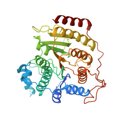
    Histone deacetylases (HDACs) are a family of enzymes involved in the regulation of gene expression, DNA repair, and stress response. These processes often are altered in tumors, and HDAC inhibitors have had pronounced antitumor activity with promising results in clinical trials. Here, we report the crystal structure of human HDAC8 in complex with a hydroxamic acid inhibitor. Such a structure of a eukaryotic zinc-dependent HDAC has not be described previously. Similar to bacterial HDAC-like protein, HDAC8 folds in a single alpha/beta domain. The inhibitor and the zinc-binding sites are similar in both proteins. However, significant differences are observed in the length and structure of the loops surrounding the active site, including the presence of two potassium ions in HDAC8 structure, one of which interacts with key catalytic residues. CD data suggest a direct role of potassium in the fold stabilization of HDAC8. Knockdown of HDAC8 by RNA interference inhibits growth of human lung, colon, and cervical cancer cell lines, highlighting the importance of this HDAC subtype for tumor cell proliferation. Our findings open the way for the design and development of selective inhibitors of HDAC8 as possible antitumor agents.


  • Organizational Affiliation

    Department of Biochemistry, Istituto di Ricerche di Biologia Molecolare P. Angeletti, 00040 Pomezia, Rome, Italy.


Macromolecules
Find similar proteins by:  (by identity cutoff)  |  3D Structure
Entity ID: 1
MoleculeChains Sequence LengthOrganismDetailsImage
HISTONE DEACETYLASE 8
A, B
377Homo sapiensMutation(s): 0 
UniProt & NIH Common Fund Data Resources
Find proteins for Q9BY41 (Homo sapiens)
Explore Q9BY41 
Go to UniProtKB:  Q9BY41
PHAROS:  Q9BY41
GTEx:  ENSG00000147099 
Entity Groups  
Sequence Clusters30% Identity50% Identity70% Identity90% Identity95% Identity100% Identity
UniProt GroupQ9BY41
Sequence Annotations
Expand
  • Reference Sequence
Small Molecules
Ligands 3 Unique
IDChains Name / Formula / InChI Key2D Diagram3D Interactions
NHB
Query on NHB

Download Ideal Coordinates CCD File 
F [auth A],
J [auth B]
N-HYDROXY-4-(METHYL{[5-(2-PYRIDINYL)-2-THIENYL]SULFONYL}AMINO)BENZAMIDE
C17 H15 N3 O4 S2
LFGYSFPVLMPUPE-UHFFFAOYSA-N
ZN
Query on ZN

Download Ideal Coordinates CCD File 
C [auth A],
G [auth B]
ZINC ION
Zn
PTFCDOFLOPIGGS-UHFFFAOYSA-N
K
Query on K

Download Ideal Coordinates CCD File 
D [auth A],
E [auth A],
H [auth B],
I [auth B]
POTASSIUM ION
K
NPYPAHLBTDXSSS-UHFFFAOYSA-N
Binding Affinity Annotations 
IDSourceBinding Affinity
NHB PDBBind:  1W22 IC50: 175.5 (nM) from 1 assay(s)
Binding MOAD:  1W22 IC50: 175.5 (nM) from 1 assay(s)
Experimental Data & Validation

Experimental Data

  • Method: X-RAY DIFFRACTION
  • Resolution: 2.50 Å
  • R-Value Free: 0.250 
  • R-Value Work: 0.210 
  • Space Group: P 1 21 1
Unit Cell:
Length ( Å )Angle ( ˚ )
a = 51.746α = 90
b = 83.532β = 97.48
c = 94.653γ = 90
Software Package:
Software NamePurpose
CNXrefinement
MOSFLMdata reduction
SCALAdata scaling
AMoREphasing

Structure Validation

View Full Validation Report


Ligand Structure Quality Assessment 


Entry History 

Deposition Data

Revision History  (Full details and data files)

  • Version 1.0: 2004-09-24
    Type: Initial release
  • Version 1.1: 2013-01-30
    Changes: Data collection, Database references, Derived calculations, Non-polymer description, Other, Refinement description, Structure summary, Version format compliance
  • Version 1.2: 2023-12-13
    Changes: Data collection, Database references, Derived calculations, Other, Refinement description