1VSC

VCAM-1


Experimental Data Snapshot

  • Method: X-RAY DIFFRACTION
  • Resolution: 1.90 Å
  • R-Value Free: 0.289 
  • R-Value Work: 0.204 
  • R-Value Observed: 0.204 

wwPDB Validation   3D Report Full Report


This is version 1.2 of the entry. See complete history


Literature

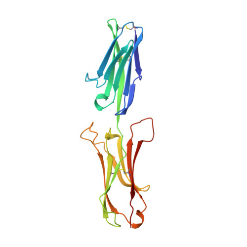
The crystal structure of an N-terminal two-domain fragment of vascular cell adhesion molecule 1 (VCAM-1): a cyclic peptide based on the domain 1 C-D loop can inhibit VCAM-1-alpha 4 integrin interaction.

Wang, J.H.Pepinsky, R.B.Stehle, T.Liu, J.H.Karpusas, M.Browning, B.Osborn, L.

(1995) Proc Natl Acad Sci U S A 92: 5714-5718

  • DOI: https://doi.org/10.1073/pnas.92.12.5714
  • Primary Citation of Related Structures:  
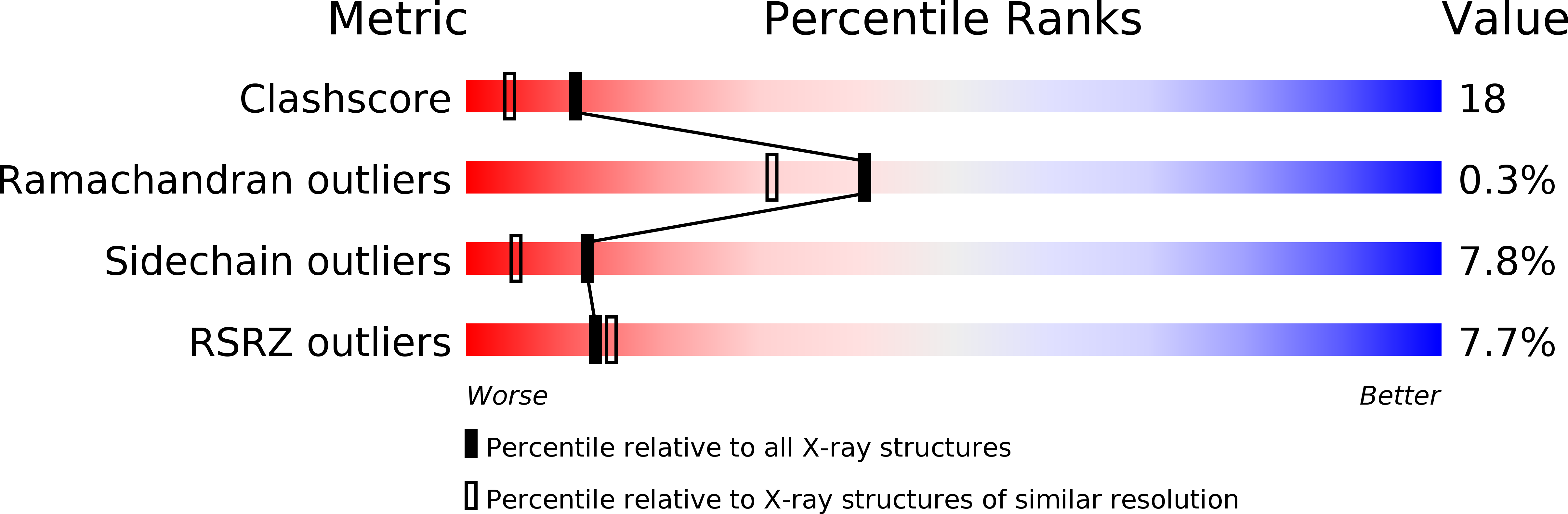
    1VSC

  • PubMed Abstract: 

    Vascular cell adhesion molecule 1 (VCAM-1) represents a structurally and functionally distinct class of immunoglobulin superfamily molecules that bind leukocyte integrins and are involved in inflammatory and immune functions. X-ray crystallography defines the three-dimensional structure of the N-terminal two-domain fragment that participates in ligand binding. Residues in domain 1 important for ligand binding reside in the C-D loop, which projects markedly from one face of the molecule near the contact between domains 1 and 2. A cyclic peptide that mimics this loop inhibits binding of alpha 4 beta 1 integrin-bearing cells to VCAM-1. These data demonstrate how crystallographic structural information can be used to design a small molecule inhibitor of biological function.


  • Organizational Affiliation

    Department of Molecular and Cellular Biology, Harvard University, Cambridge, MA 02138, USA.


Macromolecules
Find similar proteins by:  (by identity cutoff)  |  3D Structure
Entity ID: 1
MoleculeChains Sequence LengthOrganismDetailsImage
VASCULAR CELL ADHESION MOLECULE-1
A, B
196Homo sapiensMutation(s): 0 
Gene Names: VCAM-D1D2-IG
UniProt & NIH Common Fund Data Resources
Find proteins for P19320 (Homo sapiens)
Explore P19320 
Go to UniProtKB:  P19320
PHAROS:  P19320
GTEx:  ENSG00000162692 
Entity Groups  
Sequence Clusters30% Identity50% Identity70% Identity90% Identity95% Identity100% Identity
UniProt GroupP19320
Sequence Annotations
Expand
  • Reference Sequence
Experimental Data & Validation

Experimental Data

  • Method: X-RAY DIFFRACTION
  • Resolution: 1.90 Å
  • R-Value Free: 0.289 
  • R-Value Work: 0.204 
  • R-Value Observed: 0.204 
  • Space Group: C 1 2 1
Unit Cell:
Length ( Å )Angle ( ˚ )
a = 122.09α = 90
b = 48.86β = 117.42
c = 73.44γ = 90
Software Package:
Software NamePurpose
BUDDHAdata collection
X-PLORmodel building
X-PLORrefinement
BUDDHAdata reduction
X-PLORphasing

Structure Validation

View Full Validation Report


Entry History 

Deposition Data

Revision History  (Full details and data files)

  • Version 1.0: 1996-06-20
    Type: Initial release
  • Version 1.1: 2008-03-24
    Changes: Version format compliance
  • Version 1.2: 2011-07-13
    Changes: Version format compliance