


Crystal structure of Ribokinase (TM0960) from Thermotoga maritima at 2.15 A resolution
Joint Center for Structural Genomics (JCSG)To be published.
Experimental Data Snapshot
wwPDB Validation 3D Report Full Report

Entity ID: 1 | |||||
|---|---|---|---|---|---|
| Molecule | Chains | Sequence Length | Organism | Details | Image |
| ribokinase | 311 | Thermotoga maritima MSB8 | Mutation(s): 0 Gene Names: TM0960 EC: 2.7.1.15 | 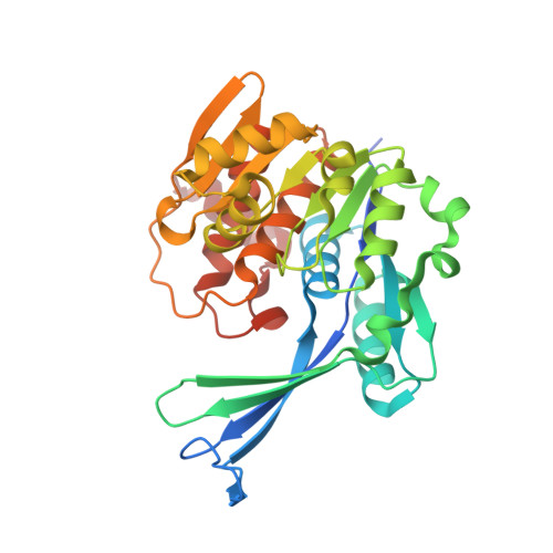 | |
UniProt | |||||
Find proteins for Q9X055 (Thermotoga maritima (strain ATCC 43589 / DSM 3109 / JCM 10099 / NBRC 100826 / MSB8)) Explore Q9X055 Go to UniProtKB: Q9X055 | |||||
Entity Groups | |||||
| Sequence Clusters | 30% Identity50% Identity70% Identity90% Identity95% Identity100% Identity | ||||
| UniProt Group | Q9X055 | ||||
Sequence AnnotationsExpand | |||||
| |||||
| Length ( Å ) | Angle ( ˚ ) |
|---|---|
| a = 181.142 | α = 90 |
| b = 45.265 | β = 93.3 |
| c = 77.26 | γ = 90 |
| Software Name | Purpose |
|---|---|
| MOSFLM | data reduction |
| SCALA | data scaling |
| MOLREP | phasing |
| REFMAC | refinement |
| CCP4 | data scaling |
RCSB PDB (citation) is hosted by
RCSB PDB is a member of the

