1VAC

MHC CLASS I H-2KB HEAVY CHAIN COMPLEXED WITH BETA-2 MICROGLOBULIN AND CHICKEN OVALBUMIN


Experimental Data Snapshot

  • Method: X-RAY DIFFRACTION
  • Resolution: 2.50 Å
  • R-Value Work: 0.173 
  • R-Value Observed: 0.173 

wwPDB Validation   3D Report Full Report


This is version 1.2 of the entry. See complete history


Literature

Crystal structure of an H-2Kb-ovalbumin peptide complex reveals the interplay of primary and secondary anchor positions in the major histocompatibility complex binding groove.

Fremont, D.H.Stura, E.A.Matsumura, M.Peterson, P.A.Wilson, I.A.

(1995) Proc Natl Acad Sci U S A 92: 2479-2483

  • DOI: https://doi.org/10.1073/pnas.92.7.2479
  • Primary Citation of Related Structures:  
    1VAC, 1VAD

  • PubMed Abstract: 

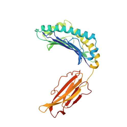
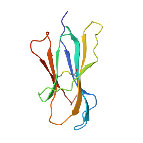
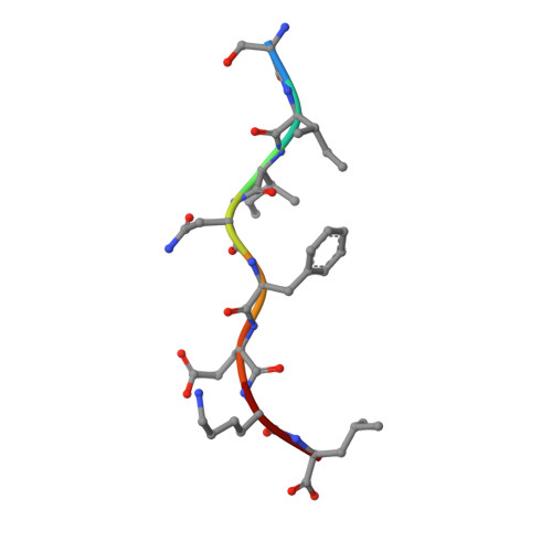
    Sequence analysis of peptides naturally presented by major histocompatibility complex (MHC) class I molecules has revealed allele-specific motifs in which the peptide length and the residues observed at certain positions are restricted. Nevertheless, peptides containing the standard motif often fail to bind with high affinity or form physiologically stable complexes. Here we present the crystal structure of a well-characterized antigenic peptide from ovalbumin [OVA-8, ovalbumin-(257-264), SIINFEKL] in complex with the murine MHC class I H-2Kb molecule at 2.5-A resolution. Hydrophobic peptide residues Ile-P2 and Phe-P5 are packed closely together into binding pockets B and C, suggesting that the interplay of peptide anchor (P5) and secondary anchor (P2) residues can couple the preferred sequences at these positions. Comparison with the crystal structures of H-2Kb in complex with peptides VSV-8 (RGYVYQGL) and SEV-9 (FAPGNYPAL), where a Tyr residue is used as the C pocket anchor, reveals that the conserved water molecule that binds into the B pocket and mediates hydrogen bonding from the buried anchor hydroxyl group could not be likewise positioned if the P2 side chain were of significant size. Based on this structural evidence, H-2Kb has at least two submotifs: one with Tyr at P5 (or P6 for nonamer peptides) and a small residue at P2 (i.e., Ala or Gly) and another with Phe at P5 and a medium-sized hydrophobic residue at P2 (i.e., Ile). Deciphering of these secondary submotifs from both crystallographic and immunological studies of MHC peptide binding should increase the accuracy of T-cell epitope prediction.


  • Organizational Affiliation

    Department of Molecular Biology, Scripps Research Institute, La Jolla, CA 92037, USA.


Macromolecules
Find similar proteins by:  (by identity cutoff)  |  3D Structure
Entity ID: 1
MoleculeChains Sequence LengthOrganismDetailsImage
MHC CLASS I H-2KB HEAVY CHAIN274Mus musculusMutation(s): 0 
Gene Names: BETA-2-MICROGLOBULIN
UniProt
Find proteins for P01901 (Mus musculus)
Explore P01901 
Go to UniProtKB:  P01901
Entity Groups  
Sequence Clusters30% Identity50% Identity70% Identity90% Identity95% Identity100% Identity
UniProt GroupP01901
Sequence Annotations
Expand
  • Reference Sequence
Find similar proteins by:  (by identity cutoff)  |  3D Structure
Entity ID: 2
MoleculeChains Sequence LengthOrganismDetailsImage
BETA-2 MICROGLOBULIN99Mus musculusMutation(s): 0 
Gene Names: BETA-2-MICROGLOBULIN
UniProt & NIH Common Fund Data Resources
Find proteins for P01887 (Mus musculus)
Explore P01887 
Go to UniProtKB:  P01887
IMPC:  MGI:88127
Entity Groups  
Sequence Clusters30% Identity50% Identity70% Identity90% Identity95% Identity100% Identity
UniProt GroupP01887
Sequence Annotations
Expand
  • Reference Sequence

Find similar proteins by:  Sequence   |   3D Structure  

Entity ID: 3
MoleculeChains Sequence LengthOrganismDetailsImage
CHICKEN OVALBUMINC [auth P]8Gallus gallusMutation(s): 0 
UniProt
Find proteins for P01012 (Gallus gallus)
Explore P01012 
Go to UniProtKB:  P01012
Entity Groups  
UniProt GroupP01012
Sequence Annotations
Expand
  • Reference Sequence
Experimental Data & Validation

Experimental Data

  • Method: X-RAY DIFFRACTION
  • Resolution: 2.50 Å
  • R-Value Work: 0.173 
  • R-Value Observed: 0.173 
  • Space Group: C 1 2 1
Unit Cell:
Length ( Å )Angle ( ˚ )
a = 118α = 90
b = 60.9β = 107.6
c = 83.8γ = 90
Software Package:
Software NamePurpose
XENGENdata collection
X-PLORmodel building
X-PLORrefinement
XENGENdata reduction
X-PLORphasing

Structure Validation

View Full Validation Report


Entry History 

Deposition Data

Revision History  (Full details and data files)

  • Version 1.0: 1996-06-20
    Type: Initial release
  • Version 1.1: 2008-03-24
    Changes: Version format compliance
  • Version 1.2: 2011-07-13
    Changes: Version format compliance