1V6P

Crystal structure of Cobrotoxin


Experimental Data Snapshot

  • Method: X-RAY DIFFRACTION
  • Resolution: 0.87 Å
  • R-Value Free: 0.153 
  • R-Value Work: 0.119 

wwPDB Validation   3D Report Full Report


This is version 1.3 of the entry. See complete history


Literature

The atomic resolution crystal structure of atratoxin determined by single wavelength anomalous diffraction phasing

Lou, X.Liu, Q.Tu, X.Wang, J.Teng, M.Niu, L.Schuller, D.J.Huang, Q.Hao, Q.

(2004) J Biol Chem 279: 39094-39104

  • DOI: https://doi.org/10.1074/jbc.M403863200
  • Primary Citation of Related Structures:  
    1V6P, 1VB0

  • PubMed Abstract: 

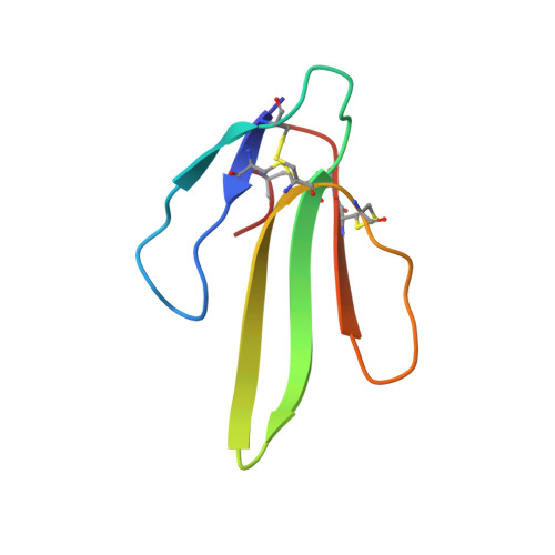
    By using single wavelength anomalous diffraction phasing based on the anomalous signal from copper atoms, the crystal structure of atratoxin was determined at the resolution of 1.5 A and was refined to an ultrahigh resolution of 0.87 A. The ultrahigh resolution electron density maps allowed the modeling of 38 amino acid residues in alternate conformations and the location of 322 of 870 possible hydrogen atoms. To get accurate information at the atomic level, atratoxin-b (an analog of atratoxin with reduced toxicity) was also refined to an atomic resolution of 0.92 A. By the sequence and structural comparison of these two atratoxins, Arg(33) and Arg(36) were identified to be critical to their varied toxicity. The effect of copper ions on the distribution of hydrogen atoms in atratoxin was discussed, and the interactions between copper ions and protein residues were analyzed based on a statistical method, revealing a novel pentahedral copper-binding motif.


  • Organizational Affiliation

    Key Laboratory of Structural Biology, Chinese Academy of Sciences, University of Science and Technology of China, 96 Jinzhai Road, Hefei, Anhui 230026, China.


Macromolecules
Find similar proteins by:  (by identity cutoff)  |  3D Structure
Entity ID: 1
MoleculeChains Sequence LengthOrganismDetailsImage
Cobrotoxin
A, B
62Naja atraMutation(s): 0 
UniProt
Find proteins for P60770 (Naja atra)
Explore P60770 
Go to UniProtKB:  P60770
Entity Groups  
Sequence Clusters30% Identity50% Identity70% Identity90% Identity95% Identity100% Identity
UniProt GroupP60770
Sequence Annotations
Expand
  • Reference Sequence
Small Molecules
Ligands 4 Unique
IDChains Name / Formula / InChI Key2D Diagram3D Interactions
CU
Query on CU

Download Ideal Coordinates CCD File 
C [auth A]
D [auth A]
E [auth A]
F [auth A]
G [auth A]
C [auth A],
D [auth A],
E [auth A],
F [auth A],
G [auth A],
H [auth A],
I [auth A],
J [auth A],
K [auth A],
L [auth A],
R [auth B],
S [auth B]
COPPER (II) ION
Cu
JPVYNHNXODAKFH-UHFFFAOYSA-N
EOH
Query on EOH

Download Ideal Coordinates CCD File 
P [auth A],
Q [auth A]
ETHANOL
C2 H6 O
LFQSCWFLJHTTHZ-UHFFFAOYSA-N
CL
Query on CL

Download Ideal Coordinates CCD File 
M [auth A]CHLORIDE ION
Cl
VEXZGXHMUGYJMC-UHFFFAOYSA-M
NA
Query on NA

Download Ideal Coordinates CCD File 
N [auth A],
O [auth A]
SODIUM ION
Na
FKNQFGJONOIPTF-UHFFFAOYSA-N
Experimental Data & Validation

Experimental Data

  • Method: X-RAY DIFFRACTION
  • Resolution: 0.87 Å
  • R-Value Free: 0.153 
  • R-Value Work: 0.119 
  • Space Group: C 2 2 21
Unit Cell:
Length ( Å )Angle ( ˚ )
a = 46.808α = 90
b = 47.287β = 90
c = 90.018γ = 90
Software Package:
Software NamePurpose
SCALEPACKdata scaling
DENZOdata reduction
CNSrefinement
SHELXmodel building
SHELXL-97refinement

Structure Validation

View Full Validation Report


Entry History 

Deposition Data

Revision History  (Full details and data files)

  • Version 1.0: 2004-12-21
    Type: Initial release
  • Version 1.1: 2008-04-27
    Changes: Version format compliance
  • Version 1.2: 2011-07-13
    Changes: Version format compliance
  • Version 1.3: 2023-12-27
    Changes: Data collection, Database references, Derived calculations