1UOU

Crystal structure of human thymidine phosphorylase in complex with a small molecule inhibitor


Experimental Data Snapshot

  • Method: X-RAY DIFFRACTION
  • Resolution: 2.11 Å
  • R-Value Free: 0.258 
  • R-Value Work: 0.188 
  • R-Value Observed: 0.192 

wwPDB Validation   3D Report Full Report


Ligand Structure Quality Assessment 


This is version 1.3 of the entry. See complete history


Literature

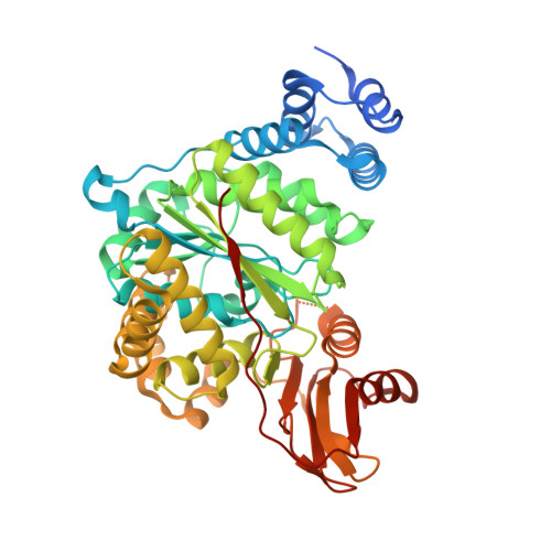
Crystal Structure of Human Thymidine Phosphorylase in Complex with a Small Molecule Inhibitor

Norman, R.A.Barry, S.T.Bate, M.Breed, J.Colls, J.G.Ernill, R.J.Luke, R.W.A.Minshull, C.A.Mcalister, M.S.B.Mccall, E.J.Mcmicken, H.H.J.Paterson, D.S.Timms, D.Tucker, J.A.Pauptit, R.A.

(2004) Structure 12: 75

  • DOI: https://doi.org/10.1016/j.str.2003.11.018
  • Primary Citation of Related Structures:  
    1UOU

  • PubMed Abstract: 

    Human thymidine phosphorylase (HTP), also known as platelet-derived endothelial cell growth factor (PD-ECGF), is overexpressed in certain solid tumors where it is linked to poor prognosis. HTP expression is utilized for certain chemotherapeutic strategies and is also thought to play a role in tumor angiogenesis. We determined the structure of HTP bound to the small molecule inhibitor 5-chloro-6-[1-(2-iminopyrrolidinyl) methyl] uracil hydrochloride (TPI). The inhibitor appears to mimic the substrate transition state, which may help explain the potency of this inhibitor and the catalytic mechanism of pyrimidine nucleotide phosphorylases (PYNPs). Further, we have confirmed the validity of the HTP structure as a template for structure-based drug design by predicting binding affinities for TPI and other known HTP inhibitors using in silico docking techniques. This work provides the first structural insight into the binding mode of any inhibitor to this important drug target and forms the basis for designing novel inhibitors for use in anticancer therapy.


  • Organizational Affiliation

    AstraZeneca, Alderley Park, Macclesfield, Cheshire SK10 4TG, UK.


Macromolecules
Find similar proteins by:  (by identity cutoff)  |  3D Structure
Entity ID: 1
MoleculeChains Sequence LengthOrganismDetailsImage
THYMIDINE PHOSPHORYLASE474Homo sapiensMutation(s): 0 
EC: 2.4.2.4
UniProt & NIH Common Fund Data Resources
Find proteins for P19971 (Homo sapiens)
Explore P19971 
Go to UniProtKB:  P19971
PHAROS:  P19971
GTEx:  ENSG00000025708 
Entity Groups  
Sequence Clusters30% Identity50% Identity70% Identity90% Identity95% Identity100% Identity
UniProt GroupP19971
Sequence Annotations
Expand
  • Reference Sequence
Small Molecules
Ligands 1 Unique
IDChains Name / Formula / InChI Key2D Diagram3D Interactions
CMU
Query on CMU

Download Ideal Coordinates CCD File 
B [auth A]5-CHLORO-6-(1-(2-IMINOPYRROLIDINYL) METHYL) URACIL
C9 H11 Cl N4 O2
QQHMKNYGKVVGCZ-WDZFZDKYSA-N
Binding Affinity Annotations 
IDSourceBinding Affinity
CMU PDBBind:  1UOU Ki: 20 (nM) from 1 assay(s)
BindingDB:  1UOU Ki: min: 1.3, max: 20 (nM) from 3 assay(s)
IC50: min: 20, max: 35 (nM) from 4 assay(s)
Binding MOAD:  1UOU Ki: 20 (nM) from 1 assay(s)
Experimental Data & Validation

Experimental Data

  • Method: X-RAY DIFFRACTION
  • Resolution: 2.11 Å
  • R-Value Free: 0.258 
  • R-Value Work: 0.188 
  • R-Value Observed: 0.192 
  • Space Group: C 1 2 1
Unit Cell:
Length ( Å )Angle ( ˚ )
a = 69.99α = 90
b = 66.09β = 106.54
c = 96.87γ = 90
Software Package:
Software NamePurpose
REFMACrefinement
MOSFLMdata reduction
SCALAdata scaling
AMoREphasing

Structure Validation

View Full Validation Report


Ligand Structure Quality Assessment 


Entry History 

Revision History  (Full details and data files)

  • Version 1.0: 2004-01-22
    Type: Initial release
  • Version 1.1: 2011-05-08
    Changes: Version format compliance
  • Version 1.2: 2011-07-13
    Changes: Version format compliance
  • Version 1.3: 2023-12-13
    Changes: Data collection, Database references, Other, Refinement description