1UNN

Complex of beta-clamp processivity factor and little finger domain of PolIV


Experimental Data Snapshot

  • Method: X-RAY DIFFRACTION
  • Resolution: 1.90 Å
  • R-Value Free: 0.240 
  • R-Value Work: 0.179 

wwPDB Validation   3D Report Full Report


This is version 1.3 of the entry. See complete history


Literature

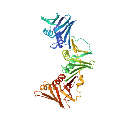
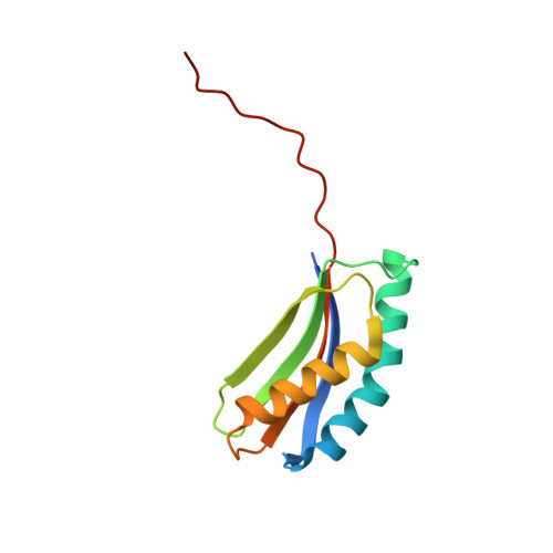
Structural Basis for Recruitment of Translesion DNA Polymerase Pol Iv/Dinb to the Beta-Clamp

Bunting, K.A.Roe, S.M.Pearl, L.H.

(2003) EMBO J 22: 5883

  • DOI: https://doi.org/10.1093/emboj/cdg568
  • Primary Citation of Related Structures:  
    1UNN

  • PubMed Abstract: 

    Y-family DNA polymerases can extend primer strands across template strand lesions that stall replicative polymerases. The poor processivity and fidelity of these enzymes, key to their biological role, requires that their access to the primer-template junction is both facilitated and regulated in order to minimize mutations. These features are believed to be provided by interaction with processivity factors, beta-clamp or proliferating cell nuclear antigen (PCNA), which are also essential for the function of replicative DNA polymerases. The basis for this interaction is revealed by the crystal structure of the complex between the 'little finger' domain of the Y-family DNA polymerase Pol IV and the beta-clamp processivity factor, both from Escherichia coli. The main interaction involves a C-terminal peptide of Pol IV, and is similar to interactions seen between isolated peptides and other processivity factors. However, this first structure of an entire domain of a binding partner with an assembled clamp reveals a substantial secondary interface, which maintains the polymerase in an inactive orientation, and may regulate the switch between replicative and Y-family DNA polymerases in response to a template strand lesion.


  • Organizational Affiliation

    The Institute of Cancer Research, Chester Beatty Laboratories, 237 Fulham Road, London SW3 6JB, UK.


Macromolecules
Find similar proteins by:  (by identity cutoff)  |  3D Structure
Entity ID: 1
MoleculeChains Sequence LengthOrganismDetailsImage
DNA POLYMERASE III BETA SUBUNIT
A, B
366Escherichia coliMutation(s): 0 
EC: 2.7.7.7
UniProt
Find proteins for P0A988 (Escherichia coli (strain K12))
Explore P0A988 
Go to UniProtKB:  P0A988
Entity Groups  
Sequence Clusters30% Identity50% Identity70% Identity90% Identity95% Identity100% Identity
UniProt GroupP0A988
Sequence Annotations
Expand
  • Reference Sequence
Find similar proteins by:  (by identity cutoff)  |  3D Structure
Entity ID: 2
MoleculeChains Sequence LengthOrganismDetailsImage
DNA POLYMERASE IV
C, D
115Escherichia coliMutation(s): 0 
EC: 2.7.7.7
UniProt
Find proteins for Q47155 (Escherichia coli (strain K12))
Explore Q47155 
Go to UniProtKB:  Q47155
Entity Groups  
Sequence Clusters30% Identity50% Identity70% Identity90% Identity95% Identity100% Identity
UniProt GroupQ47155
Sequence Annotations
Expand
  • Reference Sequence
Experimental Data & Validation

Experimental Data

  • Method: X-RAY DIFFRACTION
  • Resolution: 1.90 Å
  • R-Value Free: 0.240 
  • R-Value Work: 0.179 
  • Space Group: P 21 21 2
Unit Cell:
Length ( Å )Angle ( ˚ )
a = 146.451α = 90
b = 70.117β = 90
c = 110.916γ = 90
Software Package:
Software NamePurpose
REFMACrefinement
MOSFLMdata reduction
SCALAdata scaling
AMoREphasing

Structure Validation

View Full Validation Report


Entry History 

Deposition Data

Revision History  (Full details and data files)

  • Version 1.0: 2003-11-06
    Type: Initial release
  • Version 1.1: 2011-07-13
    Changes: Advisory, Version format compliance
  • Version 1.2: 2019-05-08
    Changes: Data collection, Experimental preparation, Other
  • Version 1.3: 2023-12-13
    Changes: Data collection, Database references, Derived calculations, Other, Refinement description