1UH9

Crystal structure of rhizopuspepsin at pH 7.0


Experimental Data Snapshot

  • Method: X-RAY DIFFRACTION
  • Resolution: 2.00 Å
  • R-Value Free: 0.194 
  • R-Value Work: 0.157 

wwPDB Validation   3D Report Full Report


This is version 1.3 of the entry. See complete history


Literature

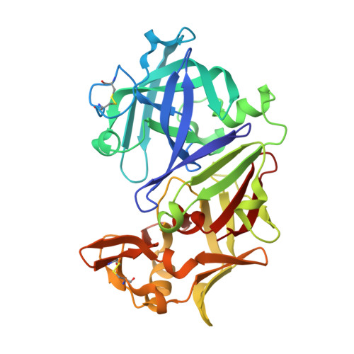
Effect of pH on the structure of rhizopuspepsin.

Prasad, B.V.Suguna, K.

(2003) Acta Crystallogr D Biol Crystallogr 59: 1755-1761

  • DOI: https://doi.org/10.1107/s0907444903016068
  • Primary Citation of Related Structures:  
    1UH7, 1UH8, 1UH9

  • PubMed Abstract: 

    The crystal structure of rhizopuspepsin has been determined at three different pH values (4.6, 7.0 and 8.0) and compared with the previously reported structure at pH 6.0. A pH-sensitive region in the protein has been identified where certain structural changes take place at pH 8.0. An increase in the mobility of loops, weakening of hydrogen bonding and ionic interactions and a change in the water structure have been observed in this region. The loop between the first and the second beta-strands of the N-terminus shows increased mobility at high pH. This loop is known to be highly flexible in aspartic proteinases, aiding in relocating the N-terminal beta-strand segment in pH-related structural transformations. The observed changes in rhizopuspepsin indicate the triggering of a possible denatured state by high pH. The conformation of the active aspartates and the geometry of the catalytic site exhibit remarkable rigidity in this pH range.


  • Organizational Affiliation

    Molecular Biophysics Unit, Indian Institute of Science, Bangalore 560 012, India.


Macromolecules
Find similar proteins by:  (by identity cutoff)  |  3D Structure
Entity ID: 1
MoleculeChains Sequence LengthOrganismDetailsImage
hizopuspepsin I325Rhizopus microsporus var. chinensisMutation(s): 0 
EC: 3.4.23.6
UniProt
Find proteins for P06026 (Rhizopus chinensis)
Explore P06026 
Go to UniProtKB:  P06026
Entity Groups  
Sequence Clusters30% Identity50% Identity70% Identity90% Identity95% Identity100% Identity
UniProt GroupP06026
Sequence Annotations
Expand
  • Reference Sequence
Experimental Data & Validation

Experimental Data

  • Method: X-RAY DIFFRACTION
  • Resolution: 2.00 Å
  • R-Value Free: 0.194 
  • R-Value Work: 0.157 
  • Space Group: P 21 21 21
Unit Cell:
Length ( Å )Angle ( ˚ )
a = 60.191α = 90
b = 60.789β = 90
c = 106.988γ = 90
Software Package:
Software NamePurpose
DENZOdata reduction
SCALEPACKdata scaling
CNSrefinement
CNSphasing

Structure Validation

View Full Validation Report


Entry History 

Deposition Data

Revision History  (Full details and data files)

  • Version 1.0: 2004-06-26
    Type: Initial release
  • Version 1.1: 2008-04-27
    Changes: Version format compliance
  • Version 1.2: 2011-07-13
    Changes: Version format compliance
  • Version 1.3: 2023-10-25
    Changes: Data collection, Database references, Refinement description