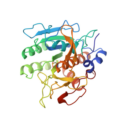
Electric fields in active sites: substrate switching from null to strong fields in thiol- and selenol-subtilisins.
Dinakarpandian, D., Shenoy, B.C., Hilvert, D., McRee, D.E., McTigue, M., Carey, P.R.(1999) Biochemistry 38: 6659-6667
- PubMed: 10350485
- DOI: https://doi.org/10.1021/bi9902541
- Primary Citation of Related Structures:
1UBN - PubMed Abstract:
Although known to be important factors in promoting catalysis, electric field effects in enzyme active sites are difficult to characterize from an experimental standpoint. Among optical probes of electric fields, Raman spectroscopy has the advantage of being able to distinguish electronic ground-state and excited-state effects. Earlier Raman studies on acyl derivatives of cysteine proteases [Doran, J. D., and Carey, P. R. (1996) Biochemistry 35, 12495-502], where the acyl group has extensive pi-electron conjugation, showed that electric field effects in the active site manifest themselves by polarizing the pi-electrons of the acyl group. Polarization gives rise to large shifts in certain Raman bands, e.g. , the C=C stretching band of the alpha,beta-unsaturated acyl group, and a large red shift in the absorption maximum. It was postulated that a major source of polarization is the alpha-helix dipole that originates from the alpha-helix terminating at the active-site cysteine of the cysteine protease family. In contrast, using the acyl group 5-methylthiophene acryloyl (5-MTA) as an active-site Raman probe, acyl enzymes of thiol- or selenol-subtilisin exhibit no polarization even though the acylating amino acid is at the terminus of an alpha-helix. Quantum mechanical calculations on 5-MTA ethyl thiol and selenol ethyl esters allowed us to identify the conformational states of these molecules along with their corresponding vibrational signatures. The Raman spectra of 5-MTA thiol and selenol subtilisins both showed that the acyl group binds in a single conformation in the active site that is s-trans about the =C-C=O single bond. Moreover, the positions of the C=C stretching bands show that the acyl group is not experiencing polarization. However, the release of steric constraints in the active site by mutagenesis, by creating the N155G form of selenol-subtilisin and the P225A form of thiol-subtilisin, results in the appearance of a second conformer in the active sites that is s-cis about the =C-C=O bond. The Raman signature of this second conformer indicates that it is strongly polarized with a permanent dipole being set up through the acyl group's pi-electron chain. Molecular modeling for 5-MTA in the active sites of selenol-subtilisin and N155G selenol-subtilisin confirms the findings from Raman spectroscopic studies and identifies the active-site features that give rise to polarization. The determinants of polarization appear to be strong electron pull at the acyl carbonyl group by a combination of hydrogen bonds and the field at the N-terminus of the alpha-helix and electron push from a negatively charged group placed at the opposite end of the chromophore.
Organizational Affiliation:
Department of Biochemistry, Case Western Reserve University, Cleveland, Ohio 44106, USA.


