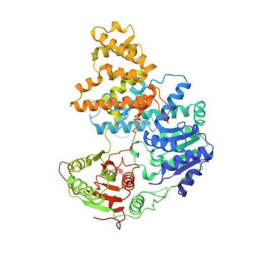
1UAA

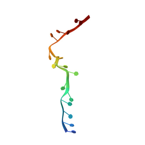
E. COLI REP HELICASE/DNA COMPLEX


Experimental Data Snapshot

  • Method: X-RAY DIFFRACTION
  • Resolution: 3.00 Å
  • R-Value Free: 0.328 
  • R-Value Work: 0.228 

wwPDB Validation   3D Report Full Report


This is version 1.3 of the entry. See complete history


Literature

Major domain swiveling revealed by the crystal structures of complexes of E. coli Rep helicase bound to single-stranded DNA and ADP.

Korolev, S.Hsieh, J.Gauss, G.H.Lohman, T.M.Waksman, G.

(1997) Cell 90: 635-647

  • DOI: https://doi.org/10.1016/s0092-8674(00)80525-5
  • Primary Citation of Related Structures:  
    1UAA

  • PubMed Abstract: 

    Crystal structures of binary and ternary complexes of the E. coli Rep helicase bound to single-stranded (ss) DNA or ssDNA and ADP were determined to a resolution of 3.0 A and 3.2 A, respectively. The asymmetric unit in the crystals contains two Rep monomers differing from each other by a large reorientation of one of the domains, corresponding to a swiveling of 130 degrees about a hinge region. Such domain movements are sufficiently large to suggest that these may be coupled to translocation of the Rep dimer along DNA. The ssDNA binding site involves the helicase motifs Ia, III, and V, whereas the ADP binding site involves helicase motifs I and IV. Residues in motifs II and VI may function to transduce the allosteric effects of nucleotides on DNA binding. These structures represent the first view of a DNA helicase bound to DNA.


  • Organizational Affiliation

    Department of Biochemistry and Molecular Biophysics, Washington University School of Medicine, St. Louis, Missouri 63110, USA.


Macromolecules

Find similar proteins by:  (by identity cutoff)  |  3D Structure
Entity ID: 2
MoleculeChains Sequence LengthOrganismDetailsImage
PROTEIN (ATP-DEPENDENT DNA HELICASE REP.)B [auth A],
C [auth B]
673Escherichia coli (strain K12)Mutation(s): 0 
Gene Names: rep
EC: 3.6.1
UniProt
Find proteins for P09980 (Escherichia coli (strain K12))
Explore P09980 
Go to UniProtKB:  P09980
Entity Groups  
Sequence Clusters30% Identity50% Identity70% Identity90% Identity95% Identity100% Identity
UniProt GroupP09980
Sequence Annotations
Expand
  • Reference Sequence

Find similar nucleic acids by:  Sequence   |   3D Structure  

Entity ID: 1
MoleculeChains LengthOrganismImage
DNA (5'-D(*TP*TP*TP*TP*TP*TP*TP*TP*TP*TP*TP*TP*TP*TP*TP*T)-3')A [auth C]16Escherichia coli
Sequence Annotations
Expand
  • Reference Sequence
Experimental Data & Validation

Experimental Data

  • Method: X-RAY DIFFRACTION
  • Resolution: 3.00 Å
  • R-Value Free: 0.328 
  • R-Value Work: 0.228 
  • Space Group: P 43 21 2
Unit Cell:
Length ( Å )Angle ( ˚ )
a = 141.8α = 90
b = 141.8β = 90
c = 284.8γ = 90
Software Package:
Software NamePurpose
DENZOdata reduction
X-PLORrefinement

Structure Validation

View Full Validation Report


Entry History 

Deposition Data

Revision History  (Full details and data files)

  • Version 1.0: 1998-07-09
    Type: Initial release
  • Version 1.1: 2008-05-22
    Changes: Version format compliance
  • Version 1.2: 2011-07-13
    Changes: Version format compliance
  • Version 1.3: 2024-02-14
    Changes: Data collection, Database references